6KPF | pdb_00006kpf

Cryo-EM structure of a class A GPCR with G protein complex


Experimental Data Snapshot

  • Method: ELECTRON MICROSCOPY
  • Resolution: 2.90 Å
  • Aggregation State: PARTICLE 
  • Reconstruction Method: SINGLE PARTICLE 

Starting Model: experimental
View more details

wwPDB Validation 3D Report Full Report

Validation slider image for 6KPF

This is version 2.1 of the entry. See complete history

Literature

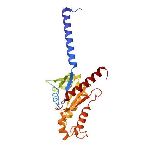
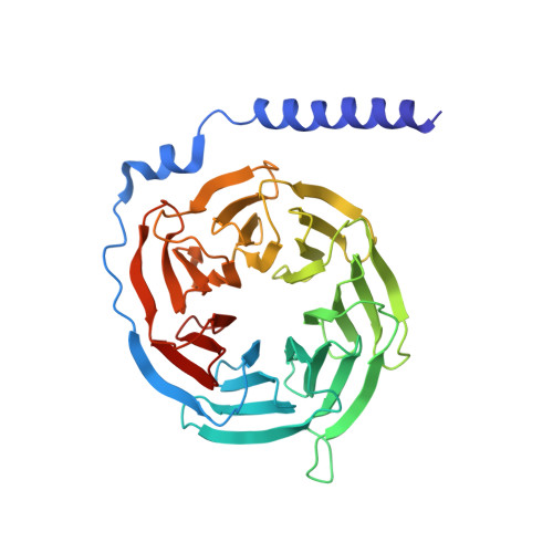
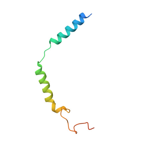
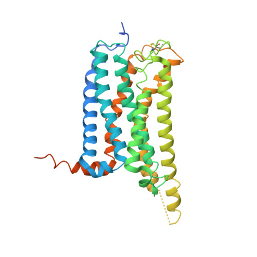
Activation and Signaling Mechanism Revealed by Cannabinoid Receptor-GiComplex Structures.

Hua, T.Li, X.Wu, L.Iliopoulos-Tsoutsouvas, C.Wang, Y.Wu, M.Shen, L.Johnston, C.A.Nikas, S.P.Song, F.Song, X.Yuan, S.Sun, Q.Wu, Y.Jiang, S.Grim, T.W.Benchama, O.Stahl, E.L.Zvonok, N.Zhao, S.Bohn, L.M.Makriyannis, A.Liu, Z.J.

(2020) Cell 180: 655

  • DOI: https://doi.org/10.1016/j.cell.2020.01.008
  • Primary Citation Related Structures: 
    6KPC, 6KPF, 6KPG

  • PubMed Abstract: 

    Human endocannabinoid systems modulate multiple physiological processes mainly through the activation of cannabinoid receptors CB1 and CB2. Their high sequence similarity, low agonist selectivity, and lack of activation and G protein-coupling knowledge have hindered the development of therapeutic applications. Importantly, missing structural information has significantly held back the development of promising CB2-selective agonist drugs for treating inflammatory and neuropathic pain without the psychoactivity of CB1. Here, we report the cryoelectron microscopy structures of synthetic cannabinoid-bound CB2 and CB1 in complex with G i , as well as agonist-bound CB2 crystal structure. Of important scientific and therapeutic benefit, our results reveal a diverse activation and signaling mechanism, the structural basis of CB2-selective agonists design, and the unexpected interaction of cholesterol with CB1, suggestive of its endogenous allosteric modulating role.


  • Organizational Affiliation
    • iHuman Institute, ShanghaiTech University, Shanghai 201210, China. Electronic address: huatian@shanghaitech.edu.cn.

Macromolecule Content 

  • Total Structure Weight: 153.6 kDa 
  • Atom Count: 8,775 
  • Modeled Residue Count: 1,135 
  • Deposited Residue Count: 1,384 
  • Unique protein chains: 5

Macromolecules

Find similar proteins by:|  3D Structure
Entity ID: 1
MoleculeChains  Sequence LengthOrganismDetailsImage
Guanine nucleotide-binding protein G(i) subunit alpha-1354Homo sapiensMutation(s): 0 
Gene Names: GNAI1
EC: 3.6.5
Membrane Entity: Yes 
UniProt & NIH Common Fund Data Resources
Find proteins for P63096 (Homo sapiens)
Explore P63096 
Go to UniProtKB:  P63096
PHAROS:  P63096
GTEx:  ENSG00000127955 
Entity Groups
Sequence Clusters30% Identity50% Identity70% Identity90% Identity95% Identity100% Identity
UniProt GroupP63096
Sequence Annotations
Expand
Reference Sequence
Find similar proteins by:|  3D Structure
Entity ID: 2
MoleculeChains  Sequence LengthOrganismDetailsImage
Guanine nucleotide-binding protein G(I)/G(S)/G(T) subunit beta-1340Homo sapiensMutation(s): 0 
Gene Names: GNB1
UniProt & NIH Common Fund Data Resources
Find proteins for P62873 (Homo sapiens)
Explore P62873 
Go to UniProtKB:  P62873
PHAROS:  P62873
GTEx:  ENSG00000078369 
Entity Groups
Sequence Clusters30% Identity50% Identity70% Identity90% Identity95% Identity100% Identity
UniProt GroupP62873
Sequence Annotations
Expand
Reference Sequence
Find similar proteins by:|  3D Structure
Entity ID: 3
MoleculeChains  Sequence LengthOrganismDetailsImage
Guanine nucleotide-binding protein G(I)/G(S)/G(O) subunit gamma-271Homo sapiensMutation(s): 0 
Gene Names: GNG2
Membrane Entity: Yes 
UniProt & NIH Common Fund Data Resources
Find proteins for P59768 (Homo sapiens)
Explore P59768 
Go to UniProtKB:  P59768
PHAROS:  P59768
GTEx:  ENSG00000186469 
Entity Groups
Sequence Clusters30% Identity50% Identity70% Identity90% Identity95% Identity100% Identity
UniProt GroupP59768
Sequence Annotations
Expand
Reference Sequence
Find similar proteins by:|  3D Structure
Entity ID: 4
MoleculeChains  Sequence LengthOrganismDetailsImage
Cannabinoid receptor 2D [auth R]360Homo sapiensMutation(s): 0 
Gene Names: CNR2CB2ACB2B
Membrane Entity: Yes 
UniProt & NIH Common Fund Data Resources
Find proteins for P34972 (Homo sapiens)
Explore P34972 
Go to UniProtKB:  P34972
PHAROS:  P34972
GTEx:  ENSG00000188822 
Entity Groups
Sequence Clusters30% Identity50% Identity70% Identity90% Identity95% Identity100% Identity
UniProt GroupP34972
Sequence Annotations
Expand
Reference Sequence
Find similar proteins by:|  3D Structure
Entity ID: 5
MoleculeChains  Sequence LengthOrganismDetailsImage
scFv16E [auth S]259Homo sapiensMutation(s): 0 

Small Molecules

Ligands 1 Unique
IDChains Name / Formula / InChI Key2D Diagram3D Interactions
E3R
(Subject of Investigation/LOI)

Query on E3R


Download:Ideal Coordinates CCD File
F [auth R]7-[(6aR,9R,10aR)-1-Hydroxy-9-(hydroxymethyl)-6,6-dimethyl-6a,7,8,9,10,10a-hexahydro-6H-benzo[c]chromen-3-yl]- 7-methyloctanenitrile
C25 H37 N O3
FTYAKKNPQHUECV-MISYRCLQSA-N

Experimental Data & Validation

Experimental Data

  • Method: ELECTRON MICROSCOPY
  • Resolution: 2.90 Å
  • Aggregation State: PARTICLE 
  • Reconstruction Method: SINGLE PARTICLE 
EM Software:
TaskSoftware PackageVersion
RECONSTRUCTIONRELION3.03
MODEL REFINEMENTPHENIX

Structure Validation

View Full Validation Report


Entry History 

Deposition Data

Revision History  (Full details and data files)

  • Version 1.0: 2020-02-12
    Type: Initial release
  • Version 1.1: 2020-03-11
    Changes: Database references
  • Version 2.0: 2022-04-27
    Changes: Advisory, Atomic model, Author supporting evidence, Data collection, Database references, Derived calculations, Non-polymer description, Source and taxonomy, Structure summary
  • Version 2.1: 2024-11-06
    Changes: Data collection, Derived calculations, Refinement description, Structure summary