


The crystal structure of cyclopenin-bound AsqJ quinary complex
Liao, H.J., Chan, N.L.(2020) J Am Chem Soc
Experimental Data Snapshot
Starting Model: experimental
View more details
(2020) J Am Chem Soc
Entity ID: 1 | |||||
|---|---|---|---|---|---|
| Molecule | Chains | Sequence Length | Organism | Details | Image |
| Iron/alpha-ketoglutarate-dependent dioxygenase asqJ | A [auth B] | 327 | Aspergillus nidulans FGSC A4 | Mutation(s): 0 Gene Names: asqJ, AN9227 EC: 1.14 (PDB Primary Data), 1.14.11.81 (UniProt) | 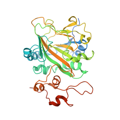 |
UniProt | |||||
Entity Groups | |||||
| Sequence Clusters | 30% Identity50% Identity70% Identity90% Identity95% Identity100% Identity | ||||
| UniProt Group | Q5AR53 | ||||
Sequence AnnotationsExpand | |||||
Reference Sequence | |||||
| Ligands 4 Unique | |||||
|---|---|---|---|---|---|
| ID | Chains | Name / Formula / InChI Key | 2D Diagram | 3D Interactions | |
| CQ9 (Subject of Investigation/LOI) Download:Ideal Coordinates CCD File | D [auth B] | (3~{R},3'~{S})-4-methyl-3'-phenyl-spiro[1~{H}-1,4-benzodiazepine-3,2'-oxirane]-2,5-dione C17 H14 N2 O3 APLKWZASYUZSBL-WMLDXEAASA-N |  | ||
| AKG Download:Ideal Coordinates CCD File | C [auth B] | 2-OXOGLUTARIC ACID C5 H6 O5 KPGXRSRHYNQIFN-UHFFFAOYSA-N |  | ||
| FE Download:Ideal Coordinates CCD File | B | FE (III) ION Fe VTLYFUHAOXGGBS-UHFFFAOYSA-N |  | ||
| PEO Download:Ideal Coordinates CCD File | E [auth B] | HYDROGEN PEROXIDE H2 O2 MHAJPDPJQMAIIY-UHFFFAOYSA-N |  | ||
| Length ( Å ) | Angle ( ˚ ) |
|---|---|
| a = 73.839 | α = 90 |
| b = 120.943 | β = 90 |
| c = 66.722 | γ = 90 |
| Software Name | Purpose |
|---|---|
| PHENIX | refinement |
| HKL-2000 | data scaling |
| PHASER | phasing |
| Coot | model building |


