6JZE

Crystal structure of VASH2-SVBP complex with the magic triangle I3C


Experimental Data Snapshot

  • Method: X-RAY DIFFRACTION
  • Resolution: 2.51 Å
  • R-Value Free: 0.266 
  • R-Value Work: 0.221 
  • R-Value Observed: 0.225 

wwPDB Validation   3D Report Full Report


Ligand Structure Quality Assessment 


This is version 2.0 of the entry. See complete history


Literature

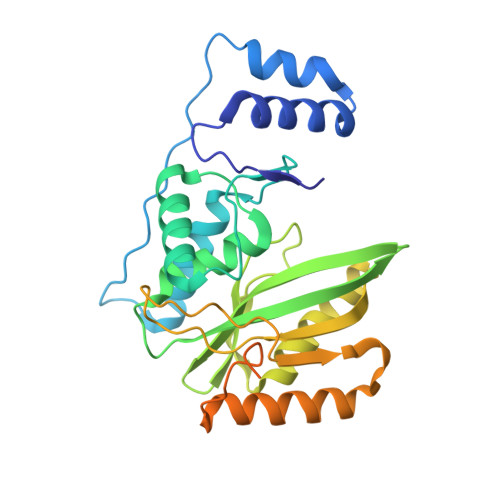
Structural basis of tubulin detyrosination by VASH2/SVBP heterodimer.

Zhou, C.Yan, L.Zhang, W.H.Liu, Z.

(2019) Nat Commun 10: 3212-3212

  • DOI: https://doi.org/10.1038/s41467-019-11277-8
  • Primary Citation of Related Structures:  
    6JZC, 6JZD, 6JZE

  • PubMed Abstract: 

    The C-terminus of α-tubulin undergoes a detyrosination/tyrosination cycle and dysregulation of this cycle is associated with cancer and other diseases. The molecular mechanisms of tubulin tyrosination are well studied, however it has remained unknown how tyrosine is cleaved from the tubulin tail. Here, we report the crystal structure of the long-sought detyrosination enzyme, the VASH2/SVBP heterodimer at 2.2 Å resolution and the structure of the tail/VASH2/SVBP complex at 2.5 Å resolution. VASH2 possesses a non-canonical Cys-His-Ser catalytic architecture for tyrosine cleavage. The dynamics of the α1- and α2- helices of VASH2 are related to the insolubility of VASH2. SVBP plays a chaperone-like role by extensively interacting with VASH2 and stabilizing these dynamic helices. A positively charged groove around the catalytic pocket and the α1- and α2- helices of VASH2 targets the tubulin tail for detyrosination. We provide insights into the mechanisms underlying the cycle of tubulin tyrosine cleavage and religation.


  • Organizational Affiliation

    National Key Laboratory of Crop Genetic Improvement, College of Life Science and Technology, Huazhong Agricultural University, Wuhan, 430070, China.


Macromolecules
Find similar proteins by:  (by identity cutoff)  |  3D Structure
Entity ID: 1
MoleculeChains Sequence LengthOrganismDetailsImage
Tubulinyl-Tyr carboxypeptidase 2309Mus musculusMutation(s): 0 
Gene Names: Vash2Vashl
EC: 3.4.17.17
UniProt
Find proteins for Q8C5G2 (Mus musculus)
Explore Q8C5G2 
Go to UniProtKB:  Q8C5G2
Entity Groups  
Sequence Clusters30% Identity50% Identity70% Identity90% Identity95% Identity100% Identity
UniProt GroupQ8C5G2
Sequence Annotations
Expand
  • Reference Sequence
Find similar proteins by:  (by identity cutoff)  |  3D Structure
Entity ID: 2
MoleculeChains Sequence LengthOrganismDetailsImage
Small vasohibin-binding protein88Homo sapiensMutation(s): 0 
Gene Names: SVBPCCDC23
UniProt & NIH Common Fund Data Resources
Find proteins for Q8N300 (Homo sapiens)
Explore Q8N300 
Go to UniProtKB:  Q8N300
PHAROS:  Q8N300
GTEx:  ENSG00000177868 
Entity Groups  
Sequence Clusters30% Identity50% Identity70% Identity90% Identity95% Identity100% Identity
UniProt GroupQ8N300
Sequence Annotations
Expand
  • Reference Sequence
Experimental Data & Validation

Experimental Data

  • Method: X-RAY DIFFRACTION
  • Resolution: 2.51 Å
  • R-Value Free: 0.266 
  • R-Value Work: 0.221 
  • R-Value Observed: 0.225 
  • Space Group: C 2 2 21
Unit Cell:
Length ( Å )Angle ( ˚ )
a = 72.69α = 90
b = 111.115β = 90
c = 126.454γ = 90
Software Package:
Software NamePurpose
PHENIXrefinement
Cootmodel building
HKL-2000data reduction
HKL-2000data scaling
SHELXDEphasing

Structure Validation

View Full Validation Report


Ligand Structure Quality Assessment 


Entry History 

Deposition Data

Revision History  (Full details and data files)

  • Version 1.0: 2019-08-07
    Type: Initial release
  • Version 2.0: 2023-04-05
    Changes: Advisory, Data collection, Database references, Derived calculations, Polymer sequence, Source and taxonomy, Structure summary