


Crystal structure of the complex of phospho pantetheine adenylyl transferase from Acinetobacter baumannii with two ascorbic acid (Vitamin-C) molecules.
Viswanathan, V., Gupta, A., Bairagya, H.R., Sharma, S., Singh, T.P.To be published.
Experimental Data Snapshot
Starting Model: experimental
View more details
Entity ID: 1 | |||||
|---|---|---|---|---|---|
| Molecule | Chains | Sequence Length | Organism | Details | Image |
| Phosphopantetheine adenylyltransferase | 163 | Acinetobacter baumannii | Mutation(s): 0 Gene Names: coaD EC: 2.7.7.3 | 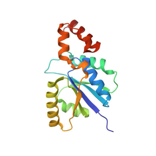 | |
UniProt | |||||
Find proteins for A0A059ZFC5 (Acinetobacter baumannii) Explore A0A059ZFC5 Go to UniProtKB: A0A059ZFC5 | |||||
Entity Groups | |||||
| Sequence Clusters | 30% Identity50% Identity70% Identity90% Identity95% Identity100% Identity | ||||
| UniProt Group | A0A059ZFC5 | ||||
Sequence AnnotationsExpand | |||||
Reference Sequence | |||||
| Ligands 2 Unique | |||||
|---|---|---|---|---|---|
| ID | Chains | Name / Formula / InChI Key | 2D Diagram | 3D Interactions | |
| ASC Download:Ideal Coordinates CCD File | B [auth A], C [auth A] | ASCORBIC ACID C6 H8 O6 CIWBSHSKHKDKBQ-JLAZNSOCSA-N |  | ||
| SO4 Download:Ideal Coordinates CCD File | D [auth A] | SULFATE ION O4 S QAOWNCQODCNURD-UHFFFAOYSA-L |  | ||
| Length ( Å ) | Angle ( ˚ ) |
|---|---|
| a = 216.667 | α = 90 |
| b = 216.667 | β = 90 |
| c = 216.667 | γ = 90 |
| Software Name | Purpose |
|---|---|
| REFMAC | refinement |
| XDS | data reduction |
| XSCALE | data scaling |
| Coot | model building |
| REFMAC | phasing |


