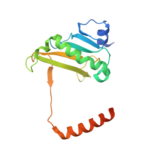
Crystal Structure of Histidine Triad Nucleotide-Binding Protein from the Pathogenic FungusCandida albicans.
Jung, A., Yun, J.S., Kim, S., Kim, S.R., Shin, M., Cho, D.H., Choi, K.S., Chang, J.H.(2019) Mol Cells 42: 56-66
- PubMed: 30622225 Search on PubMedSearch on PubMed Central
- DOI: https://doi.org/10.14348/molcells.2018.0377
- Primary Citation Related Structures:
6IQ1 - PubMed Abstract:
Histidine triad nucleotide-binding protein (HINT) is a member of the histidine triad (HIT) superfamily, which has hydrolase activity owing to a histidine triad motif. The HIT superfamily can be divided to five classes with functions in galactose metabolism, DNA repair, and tumor suppression. HINTs are highly conserved from archaea to humans and function as tumor suppressors, translation regulators, and neuropathy inhibitors. Although the structures of HINT proteins from various species have been reported, limited structural information is available for fungal species. Here, to elucidate the structural features and functional diversity of HINTs, we determined the crystal structure of HINT from the pathogenic fungus Candida albicans (CaHINT) in complex with zinc ions at a resolution of 2.5 Å. Based on structural comparisons, the monomer of CaHINT overlaid best with HINT protein from the protozoal species Leishmania major . Additionally, structural comparisons with human HINT revealed an additional helix at the C-terminus of CaHINT. Interestingly, the extended C-terminal helix interacted with the N-terminal loop (α1-β1) and with the α3 helix, which appeared to stabilize the dimerization of CaHINT. In the C-terminal region, structural and sequence comparisons showed strong relationships among 19 diverse species from archea to humans, suggesting early separation in the course of evolution. Further studies are required to address the functional significance of variations in the C-terminal region. This structural analysis of CaHINT provided important insights into the molecular aspects of evolution within the HIT superfamily.
- Department of Biology Education, Kyungpook National University, Daegu 41566, Korea.
Organizational Affiliation: