6IIH

crystal structure of mitochondrial calcium uptake 2(MICU2)


Experimental Data Snapshot

  • Method: X-RAY DIFFRACTION
  • Resolution: 1.96 Å
  • R-Value Free: 0.227 
  • R-Value Work: 0.180 
  • R-Value Observed: 0.181 

wwPDB Validation   3D Report Full Report


This is version 1.2 of the entry. See complete history


Literature

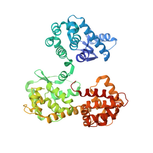
The crystal structure of MICU2 provides insight into Ca2+binding and MICU1-MICU2 heterodimer formation.

Wu, W.Shen, Q.Lei, Z.Qiu, Z.Li, D.Pei, H.Zheng, J.Jia, Z.

(2019) EMBO Rep 20: e47488-e47488

  • DOI: https://doi.org/10.15252/embr.201847488
  • Primary Citation of Related Structures:  
    6IIH

  • PubMed Abstract: 

    The mitochondrial calcium uniporter (MCU) complex mediates the uptake of Ca 2+ into mitochondria. Its activity is regulated by a heterodimer of MICU1 and MICU2, two EF-hand-containing proteins that act as the main gatekeeper of the uniporter. Herein we report the crystal structure of human MICU2 at 1.96 Å resolution. Our structure reveals a dimeric architecture of MICU2, in which each monomer adopts the canonical two-lobe structure with a pair of EF-hands in each lobe. Both Ca 2+ -bound and Ca 2+ -free EF-hands are observed in our structure. Moreover, we characterize the interaction sites within the MICU2 homodimer, as well as the MICU1-MICU2 heterodimer in both Ca 2+ -free and Ca 2+ -bound conditions. Glu242 in MICU1 and Arg352 in MICU2 are crucial for apo heterodimer formation, while Phe383 in MICU1 and Glu196 in MICU2 significantly contribute to the interaction in the Ca 2+ -bound state. Based on our structural and biochemical analyses, we propose a model for MICU1-MICU2 heterodimer formation and its conformational transition from apo to a more compact Ca 2+ -bound state, which expands our understanding of this co-regulatory mechanism critical for MCU's mitochondrial calcium uptake function.


  • Organizational Affiliation

    College of Chemistry, Beijing Normal University, Beijing, China.


Macromolecules
Find similar proteins by:  (by identity cutoff)  |  3D Structure
Entity ID: 1
MoleculeChains Sequence LengthOrganismDetailsImage
Endolysin,Calcium uptake protein 2, mitochondrial
A, B
493Tequatrovirus T4Homo sapiens
This entity is chimeric
Mutation(s): 2 
EC: 3.2.1.17
UniProt & NIH Common Fund Data Resources
Find proteins for Q8IYU8 (Homo sapiens)
Explore Q8IYU8 
Go to UniProtKB:  Q8IYU8
PHAROS:  Q8IYU8
GTEx:  ENSG00000165487 
Find proteins for P00720 (Enterobacteria phage T4)
Explore P00720 
Go to UniProtKB:  P00720
Entity Groups  
Sequence Clusters30% Identity50% Identity70% Identity90% Identity95% Identity100% Identity
UniProt GroupsP00720Q8IYU8
Sequence Annotations
Expand
  • Reference Sequence
Experimental Data & Validation

Experimental Data

  • Method: X-RAY DIFFRACTION
  • Resolution: 1.96 Å
  • R-Value Free: 0.227 
  • R-Value Work: 0.180 
  • R-Value Observed: 0.181 
  • Space Group: P 1 21 1
Unit Cell:
Length ( Å )Angle ( ˚ )
a = 66.743α = 90
b = 96.311β = 98.98
c = 85.488γ = 90
Software Package:
Software NamePurpose
PHENIXrefinement
HKL-2000data reduction
HKL-2000data scaling
PHENIXphasing

Structure Validation

View Full Validation Report


Entry History & Funding Information

Deposition Data


Funding OrganizationLocationGrant Number
National Natural Science Foundation of ChinaChina21773014

Revision History  (Full details and data files)

  • Version 1.0: 2019-08-14
    Type: Initial release
  • Version 1.1: 2019-12-18
    Changes: Database references
  • Version 1.2: 2024-03-27
    Changes: Data collection, Database references, Derived calculations