6IF1

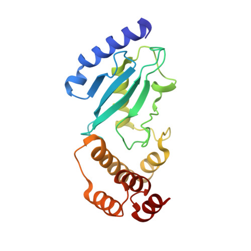
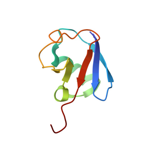
Crystal structure of Ube2K and K48-linked di-ubiquitin complex


Experimental Data Snapshot

  • Method: X-RAY DIFFRACTION
  • Resolution: 2.47 Å
  • R-Value Free: 0.236 
  • R-Value Work: 0.215 
  • R-Value Observed: 0.216 

wwPDB Validation   3D Report Full Report


This is version 1.1 of the entry. See complete history


Literature

Crystal structure of the Ube2K/E2-25K and K48-linked di-ubiquitin complex provides structural insight into the mechanism of K48-specific ubiquitin chain synthesis.

Lee, J.G.Youn, H.S.Kang, J.Y.Park, S.Y.Kidera, A.Yoo, Y.J.Eom, S.H.

(2018) Biochem Biophys Res Commun 506: 102-107

  • DOI: https://doi.org/10.1016/j.bbrc.2018.10.067
  • Primary Citation of Related Structures:  
    6IF1

  • PubMed Abstract: 

    Ubiquitin-conjugating enzymes (E2) form thioester bonds with ubiquitin (Ub), which are subsequently transferred to target proteins for cellular progress. Ube2K/E2-25K (a class II E2 enzyme) contains a C-terminal ubiquitin-associated (UBA) domain that has been suggested to control ubiquitin recognition, dimerization, or poly-ubiquitin chain formation. Ube2K is a special E2 because it synthesizes K48-linked poly-ubiquitin chains without E3 ubiquitin ligase. We found that a novel interaction between the acceptor di-Ub (Ub 2 ) and the auxiliary Ube2K promotes the discharging reaction and production of tri-Ub (Ub 3 ), probably by guiding and positioning the K48 (in the distal Ub) of the acceptor Ub 2 in the active site. We also determined the crystal structure of Ube2K-Ub 2 at 2.47 Å resolution. Based on our structural and biochemical data, we proposed a structural model of Ub 3 synthesis by Ube2K without E3.


  • Organizational Affiliation

    School of Life Sciences, Gwangju Institute of Science and Technology, Gwangju, 61005, Republic of Korea; Steitz Center for Structural Biology, Gwangju Institute of Science and Technology, Gwangju, 61005, Republic of Korea.


Macromolecules
Find similar proteins by:  (by identity cutoff)  |  3D Structure
Entity ID: 1
MoleculeChains Sequence LengthOrganismDetailsImage
Ubiquitin-conjugating enzyme E2 K
A, B
199Homo sapiensMutation(s): 0 
Gene Names: UBE2KHIP2LIG
EC: 2.3.2.23
UniProt & NIH Common Fund Data Resources
Find proteins for P61086 (Homo sapiens)
Explore P61086 
Go to UniProtKB:  P61086
PHAROS:  P61086
GTEx:  ENSG00000078140 
Entity Groups  
Sequence Clusters30% Identity50% Identity70% Identity90% Identity95% Identity100% Identity
UniProt GroupP61086
Sequence Annotations
Expand
  • Reference Sequence
Find similar proteins by:  (by identity cutoff)  |  3D Structure
Entity ID: 2
MoleculeChains Sequence LengthOrganismDetailsImage
Ubiquitin
C, D
76Homo sapiensMutation(s): 0 
Gene Names: UBB
UniProt & NIH Common Fund Data Resources
Find proteins for P0CG47 (Homo sapiens)
Explore P0CG47 
Go to UniProtKB:  P0CG47
PHAROS:  P0CG47
GTEx:  ENSG00000170315 
Entity Groups  
Sequence Clusters30% Identity50% Identity70% Identity90% Identity95% Identity100% Identity
UniProt GroupP0CG47
Sequence Annotations
Expand
  • Reference Sequence
Experimental Data & Validation

Experimental Data

  • Method: X-RAY DIFFRACTION
  • Resolution: 2.47 Å
  • R-Value Free: 0.236 
  • R-Value Work: 0.215 
  • R-Value Observed: 0.216 
  • Space Group: P 1 21 1
Unit Cell:
Length ( Å )Angle ( ˚ )
a = 41.559α = 90
b = 38.792β = 90.1
c = 190.321γ = 90
Software Package:
Software NamePurpose
PHENIXrefinement
HKL-2000data reduction
HKL-2000data scaling
PHASERphasing

Structure Validation

View Full Validation Report


Entry History 

Deposition Data

Revision History  (Full details and data files)

  • Version 1.0: 2018-11-21
    Type: Initial release
  • Version 1.1: 2023-11-22
    Changes: Data collection, Database references, Refinement description