6HZL

Crystal structure of redox-inhibited phosphoribulokinase from Synechococcus sp. (strain PCC 6301), osmate derivative


Experimental Data Snapshot

  • Method: X-RAY DIFFRACTION
  • Resolution: 2.77 Å
  • R-Value Free: 0.259 
  • R-Value Work: 0.235 
  • R-Value Observed: 0.236 

wwPDB Validation   3D Report Full Report


This is version 1.2 of the entry. See complete history


Literature

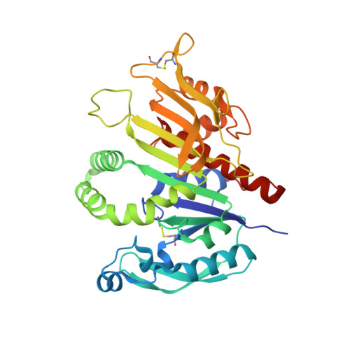
Crystal structure of phosphoribulokinase from Synechococcus sp. strain PCC 6301.

Wilson, R.H.Hayer-Hartl, M.Bracher, A.

(2019) Acta Crystallogr F Struct Biol Commun 75: 278-289

  • DOI: https://doi.org/10.1107/S2053230X19002693
  • Primary Citation of Related Structures:  
    6HZK, 6HZL

  • PubMed Abstract: 

    Phosphoribulokinase (PRK) catalyses the ATP-dependent phosphorylation of ribulose 5-phosphate to give ribulose 1,5-bisphosphate. Regulation of this reaction in response to light controls carbon fixation during photosynthesis. Here, the crystal structure of PRK from the cyanobacterium Synechococcus sp. strain PCC 6301 is presented. The enzyme is dimeric and has an α/β-fold with an 18-stranded β-sheet at its core. Interestingly, a disulfide bond is found between Cys40 and the P-loop residue Cys18, revealing the structural basis for the redox inactivation of PRK activity. A second disulfide bond appears to rigidify the dimer interface and may thereby contribute to regulation by the adaptor protein CP12 and glyceraldehyde-3-phosphate dehydrogenase.


  • Organizational Affiliation

    Department of Cellular Biochemistry, Max Planck Institute of Biochemistry, Am Klopferspitz 18, 82152 Martinsried, Germany.


Macromolecules
Find similar proteins by:  (by identity cutoff)  |  3D Structure
Entity ID: 1
MoleculeChains Sequence LengthOrganismDetailsImage
Phosphoribulokinase
A, B
333Synechococcus elongatus PCC 6301Mutation(s): 0 
Gene Names: prksyc0567_d
EC: 2.7.1.19
UniProt
Find proteins for A0A0H3K6J7 (Synechococcus sp. (strain ATCC 27144 / PCC 6301 / SAUG 1402/1))
Explore A0A0H3K6J7 
Go to UniProtKB:  A0A0H3K6J7
Entity Groups  
Sequence Clusters30% Identity50% Identity70% Identity90% Identity95% Identity100% Identity
UniProt GroupA0A0H3K6J7
Sequence Annotations
Expand
  • Reference Sequence
Experimental Data & Validation

Experimental Data

  • Method: X-RAY DIFFRACTION
  • Resolution: 2.77 Å
  • R-Value Free: 0.259 
  • R-Value Work: 0.235 
  • R-Value Observed: 0.236 
  • Space Group: H 3 2
Unit Cell:
Length ( Å )Angle ( ˚ )
a = 141.432α = 90
b = 141.432β = 90
c = 206.563γ = 120
Software Package:
Software NamePurpose
REFMACrefinement
XDSdata reduction
Aimlessdata scaling
HKL2Mapphasing
PDB_EXTRACTdata extraction

Structure Validation

View Full Validation Report


Entry History 

Deposition Data

Revision History  (Full details and data files)

  • Version 1.0: 2019-03-27
    Type: Initial release
  • Version 1.1: 2019-04-10
    Changes: Data collection, Database references
  • Version 1.2: 2019-04-17
    Changes: Data collection, Database references