


Apo structure of TP domain from Burkholderia pseudomallei penicillin-binding protein 3
Bellini, D., Koekemoer, L., Newman, H., Dowson, C.G.To be published.
Experimental Data Snapshot
Starting Model: experimental
View more details
wwPDB Validation 3D Report Full Report

Entity ID: 1 | |||||
|---|---|---|---|---|---|
| Molecule | Chains | Sequence Length | Organism | Details | Image |
| Peptidoglycan D,D-transpeptidase FtsI | 374 | Burkholderia pseudomallei | Mutation(s): 0 Gene Names: ftsI, CXQ84_03210, DP46_2053 EC: 3.4.16.4 | 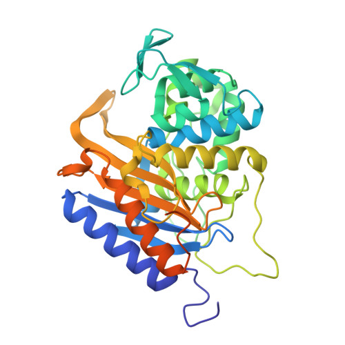 | |
UniProt | |||||
Entity Groups | |||||
| Sequence Clusters | 30% Identity50% Identity70% Identity90% Identity95% Identity100% Identity | ||||
| UniProt Group | Q63QJ1 | ||||
Sequence AnnotationsExpand | |||||
Reference Sequence | |||||
| Length ( Å ) | Angle ( ˚ ) |
|---|---|
| a = 106.394 | α = 90 |
| b = 106.394 | β = 90 |
| c = 95.678 | γ = 90 |
| Software Name | Purpose |
|---|---|
| REFMAC | refinement |
| DIALS | data reduction |
| Aimless | data scaling |
| MrBUMP | phasing |
| Funding Organization | Location | Grant Number |
|---|---|---|
| Medical Research Council (United Kingdom) | United Kingdom | grant.MR/P007503/1 |


