6HQ8 | pdb_00006hq8

Bacterial beta-1,3-oligosaccharide phosphorylase from GH149 with laminarihexaose bound at a surface site


Experimental Data Snapshot

  • Method: X-RAY DIFFRACTION
  • Resolution: 2.25 Å
  • R-Value Free: 
    0.217 (Depositor), 0.219 (DCC) 
  • R-Value Work: 
    0.180 (Depositor), 0.184 (DCC) 
  • R-Value Observed: 
    0.182 (Depositor) 

Starting Model: experimental
View more details

wwPDB Validation 3D Report Full Report

Validation slider image for 6HQ8

Ligand Structure Quality Assessment 


This is version 2.1 of the entry. See complete history

Literature

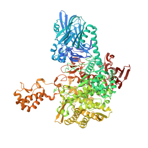
The structure of a GH149 beta-(1 → 3) glucan phosphorylase reveals a new surface oligosaccharide binding site and additional domains that are absent in the disaccharide-specific GH94 glucose-beta-(1 → 3)-glucose (laminaribiose) phosphorylase.

Kuhaudomlarp, S.Stevenson, C.E.M.Lawson, D.M.Field, R.A.

(2019) Proteins 87: 885-892

  • DOI: https://doi.org/10.1002/prot.25745
  • Primary Citation Related Structures: 
    6HQ6, 6HQ8

  • PubMed Abstract: 

    Glycoside phosphorylases (GPs) with specificity for β-(1 → 3)-gluco-oligosaccharides are potential candidate biocatalysts for oligosaccharide synthesis. GPs with this linkage specificity are found in two families thus far-glycoside hydrolase family 94 (GH94) and the recently discovered glycoside hydrolase family 149 (GH149). Previously, we reported a crystallographic study of a GH94 laminaribiose phosphorylase with specificity for disaccharides, providing insight into the enzyme's ability to recognize its' sugar substrate/product. In contrast to GH94, characterized GH149 enzymes were shown to have more flexible chain length specificity, with preference for substrate/product with higher degree of polymerization. In order to advance understanding of the specificity of GH149 enzymes, we herein solved X-ray crystallographic structures of GH149 enzyme Pro_7066 in the absence of substrate and in complex with laminarihexaose (G6). The overall domain organization of Pro_7066 is very similar to that of GH94 family enzymes. However, two additional domains flanking its catalytic domain were found only in the GH149 enzyme. Unexpectedly, the G6 complex structure revealed an oligosaccharide surface binding site remote from the catalytic site, which, we suggest, may be associated with substrate targeting. As such, this study reports the first structure of a GH149 phosphorylase enzyme acting on β-(1 → 3)-gluco-oligosaccharides and identifies structural elements that may be involved in defining the specificity of the GH149 enzymes.


  • Organizational Affiliation
    • Department of Biological Chemistry, John Innes Centre, Norwich Research Park, Norwich, UK.

Macromolecule Content 

  • Total Structure Weight: 271.87 kDa 
  • Atom Count: 18,945 
  • Modeled Residue Count: 2,276 
  • Deposited Residue Count: 2,350 
  • Unique protein chains: 1

Macromolecules

Find similar proteins by:|  3D Structure
Entity ID: 1
MoleculeChains  Sequence LengthOrganismDetailsImage
Beta-1,3-oligosaccharide phosphorylase
A, B
1,175metagenomeMutation(s): 0 
EC: 2.4.1

Oligosaccharides

Help  
Entity ID: 2
MoleculeChains Length2D Diagram GlycosylationD Interactions
beta-D-glucopyranose-(1-3)-beta-D-glucopyranose-(1-3)-beta-D-glucopyranose-(1-3)-beta-D-glucopyranose-(1-3)-beta-D-glucopyranose-(1-3)-beta-D-glucopyranose
C, D
6N/A
Glycosylation Resources
GlyTouCan: G41202OE
GlyCosmos: G41202OE
GlyGen: G41202OE

Small Molecules

Ligands 4 Unique
IDChains Name / Formula / InChI Key2D Diagram3D Interactions
BCN

Query on BCN


Download:Ideal Coordinates CCD File
G [auth A],
T [auth B]
BICINE
C6 H13 N O4
FSVCELGFZIQNCK-UHFFFAOYSA-N
SO4

Query on SO4


Download:Ideal Coordinates CCD File
E [auth A],
F [auth A],
R [auth B],
S [auth B]
SULFATE ION
O4 S
QAOWNCQODCNURD-UHFFFAOYSA-L
EDO

Query on EDO


Download:Ideal Coordinates CCD File
H [auth A]
I [auth A]
J [auth A]
K [auth A]
L [auth A]
H [auth A],
I [auth A],
J [auth A],
K [auth A],
L [auth A],
M [auth A],
N [auth A],
O [auth A],
P [auth A],
U [auth B],
V [auth B]
1,2-ETHANEDIOL
C2 H6 O2
LYCAIKOWRPUZTN-UHFFFAOYSA-N
CL

Query on CL


Download:Ideal Coordinates CCD File
Q [auth A],
W [auth B]
CHLORIDE ION
Cl
VEXZGXHMUGYJMC-UHFFFAOYSA-M

Experimental Data & Validation

Experimental Data

  • Method: X-RAY DIFFRACTION
  • Resolution: 2.25 Å
  • R-Value Free:  0.217 (Depositor), 0.219 (DCC) 
  • R-Value Work:  0.180 (Depositor), 0.184 (DCC) 
  • R-Value Observed: 0.182 (Depositor) 
Space Group: P 21 21 21
Unit Cell:
Length ( Å )Angle ( ˚ )
a = 99.129α = 90
b = 158.756β = 90
c = 178.891γ = 90
Software Package:
Software NamePurpose
XDSdata reduction
Aimlessdata scaling
REFMACrefinement
PDB_EXTRACTdata extraction
REFMACphasing

Structure Validation

View Full Validation Report


Ligand Structure Quality Assessment 


Entry History 

& Funding Information

Deposition Data


Funding OrganizationLocationGrant Number
Biotechnology and Biological Sciences Research CouncilUnited KingdomBB/J004561/1
Engineering and Physical Sciences Research CouncilUnited KingdomBB/M02903411

Revision History  (Full details and data files)

  • Version 1.0: 2019-06-12
    Type: Initial release
  • Version 1.1: 2019-09-11
    Changes: Data collection, Database references
  • Version 2.0: 2020-07-29
    Type: Remediation
    Reason: Carbohydrate remediation
    Changes: Atomic model, Data collection, Derived calculations, Structure summary
  • Version 2.1: 2024-01-24
    Changes: Data collection, Database references, Derived calculations, Refinement description, Structure summary