6HKG

Structure of FISW84 Fab Fragment

  • Classification: IMMUNE SYSTEM
  • Organism(s): Homo sapiens
  • Expression System: Homo sapiens
  • Mutation(s): No 

  • Deposited: 2018-09-06 Released: 2018-09-26 
  • Deposition Author(s): Benton, D.J., Rosenthal, P.B.
  • Funding Organization(s): Cancer Research UK, Medical Research Council (United Kingdom), Wellcome Trust, European Research Council

Experimental Data Snapshot

  • Method: X-RAY DIFFRACTION
  • Resolution: 1.87 Å
  • R-Value Free: 0.200 
  • R-Value Work: 0.183 
  • R-Value Observed: 0.183 

wwPDB Validation   3D Report Full Report


This is version 1.2 of the entry. See complete history


Literature

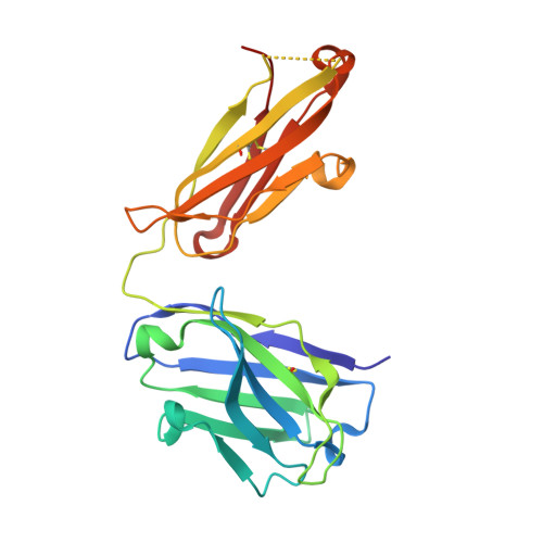
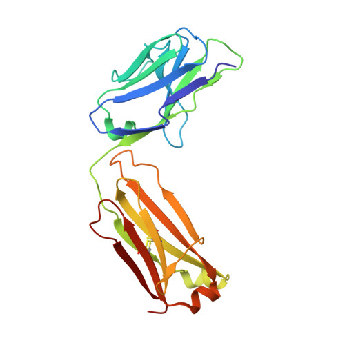
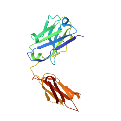
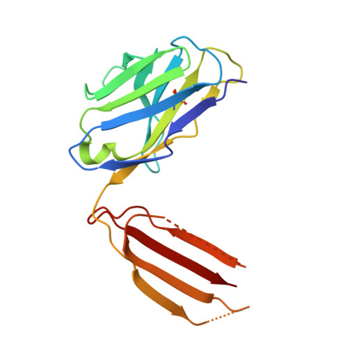
Influenza hemagglutinin membrane anchor.

Benton, D.J.Nans, A.Calder, L.J.Turner, J.Neu, U.Lin, Y.P.Ketelaars, E.Kallewaard, N.L.Corti, D.Lanzavecchia, A.Gamblin, S.J.Rosenthal, P.B.Skehel, J.J.

(2018) Proc Natl Acad Sci U S A 115: 10112-10117

  • DOI: https://doi.org/10.1073/pnas.1810927115
  • Primary Citation of Related Structures:  
    6HJN, 6HJP, 6HJQ, 6HJR, 6HKG

  • PubMed Abstract: 

    Viruses with membranes fuse them with cellular membranes, to transfer their genomes into cells at the beginning of infection. For Influenza virus, the membrane glycoprotein involved in fusion is the hemagglutinin (HA), the 3D structure of which is known from X-ray crystallographic studies. The soluble ectodomain fragments used in these studies lacked the "membrane anchor" portion of the molecule. Since this region has a role in membrane fusion, we have determined its structure by analyzing the intact, full-length molecule in a detergent micelle, using cryo-EM. We have also compared the structures of full-length HA-detergent micelles with full-length HA-Fab complex detergent micelles, to describe an infectivity-neutralizing monoclonal Fab that binds near the ectodomain membrane anchor junction. We determine a high-resolution HA structure which compares favorably in detail with the structure of the ectodomain seen by X-ray crystallography; we detect, clearly, all five carbohydrate side chains of HA; and we find that the ectodomain is joined to the membrane anchor by flexible, eight-residue-long, linkers. The linkers extend into the detergent micelle to join a central triple-helical structure that is a major component of the membrane anchor.


  • Organizational Affiliation

    Structural Biology of Disease Processes Laboratory, Francis Crick Institute, NW1 1AT London, United Kingdom; Donald.Benton@crick.ac.uk Peter.Rosenthal@crick.ac.uk John.Skehel@crick.ac.uk.


Macromolecules
Find similar proteins by:  (by identity cutoff)  |  3D Structure
Entity ID: 1
MoleculeChains Sequence LengthOrganismDetailsImage
FISW84 Fab Fragment Heavy Chain217Homo sapiensMutation(s): 0 
Entity Groups  
Sequence Clusters30% Identity50% Identity70% Identity90% Identity95% Identity100% Identity
Sequence Annotations
Expand
  • Reference Sequence
Find similar proteins by:  (by identity cutoff)  |  3D Structure
Entity ID: 2
MoleculeChains Sequence LengthOrganismDetailsImage
FISW84 Fab Fragment Light Chain214Homo sapiensMutation(s): 0 
Entity Groups  
Sequence Clusters30% Identity50% Identity70% Identity90% Identity95% Identity100% Identity
Sequence Annotations
Expand
  • Reference Sequence
Find similar proteins by:  (by identity cutoff)  |  3D Structure
Entity ID: 3
MoleculeChains Sequence LengthOrganismDetailsImage
FISW84 Fab Fragment Heavy Chain194Homo sapiensMutation(s): 0 
Entity Groups  
Sequence Clusters30% Identity50% Identity70% Identity90% Identity95% Identity100% Identity
Sequence Annotations
Expand
  • Reference Sequence
Find similar proteins by:  (by identity cutoff)  |  3D Structure
Entity ID: 4
MoleculeChains Sequence LengthOrganismDetailsImage
FISW84 Fab Fragment Light Chain159Homo sapiensMutation(s): 0 
Entity Groups  
Sequence Clusters30% Identity50% Identity70% Identity90% Identity95% Identity100% Identity
Sequence Annotations
Expand
  • Reference Sequence
Experimental Data & Validation

Experimental Data

  • Method: X-RAY DIFFRACTION
  • Resolution: 1.87 Å
  • R-Value Free: 0.200 
  • R-Value Work: 0.183 
  • R-Value Observed: 0.183 
  • Space Group: P 43 21 2
Unit Cell:
Length ( Å )Angle ( ˚ )
a = 130.16α = 90
b = 130.16β = 90
c = 167.24γ = 90
Software Package:
Software NamePurpose
PHENIXrefinement
XDSdata reduction
XSCALEdata scaling
PHASERphasing

Structure Validation

View Full Validation Report


Entry History & Funding Information

Deposition Data


Funding OrganizationLocationGrant Number
Cancer Research UKUnited KingdomFC001078
Cancer Research UKUnited KingdomFC001143
Medical Research Council (United Kingdom)United KingdomFC001078
Medical Research Council (United Kingdom)United KingdomFC001143
Wellcome TrustUnited KingdomFC001078
Wellcome TrustUnited KingdomFC001143
European Research CouncilUnited Kingdom629829

Revision History  (Full details and data files)

  • Version 1.0: 2018-09-26
    Type: Initial release
  • Version 1.1: 2018-10-10
    Changes: Data collection, Database references, Structure summary
  • Version 1.2: 2024-01-17
    Changes: Data collection, Database references, Refinement description, Structure summary