6HEF | pdb_00006hef

Room temperature structure of the (SR)Ca2+-ATPase Ca2-E1-CaAMPPCP form


Experimental Data Snapshot

  • Method: X-RAY DIFFRACTION
  • Resolution: 3.54 Å
  • R-Value Free: 
    0.257 (Depositor), 0.256 (DCC) 
  • R-Value Work: 
    0.223 (Depositor), 0.224 (DCC) 
  • R-Value Observed: 
    0.224 (Depositor) 

Starting Model: experimental
View more details

wwPDB Validation 3D Report Full Report

Validation slider image for 6HEF

Ligand Structure Quality Assessment 


This is version 1.2 of the entry. See complete history

Literature

Membrane-protein crystals for neutron diffraction.

Sorensen, T.L.M.Hjorth-Jensen, S.J.Oksanen, E.Andersen, J.L.Olesen, C.Moller, J.V.Nissen, P.

(2018) Acta Crystallogr D Struct Biol 74: 1208-1218

  • DOI: https://doi.org/10.1107/S2059798318012561
  • Primary Citation Related Structures: 
    6HEF

  • PubMed Abstract: 

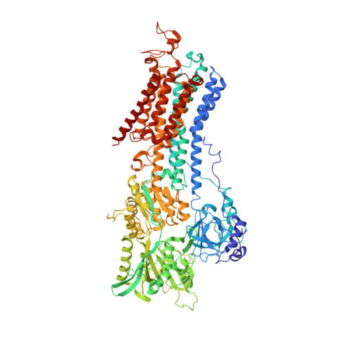
    Neutron macromolecular crystallography (NMX) has the potential to provide the experimental input to address unresolved aspects of transport mechanisms and protonation in membrane proteins. However, despite this clear scientific motivation, the practical challenges of obtaining crystals that are large enough to make NMX feasible have so far been prohibitive. Here, the potential impact on feasibility of a more powerful neutron source is reviewed and a strategy for obtaining larger crystals is formulated, exemplified by the calcium-transporting ATPase SERCA1. The challenges encountered at the various steps in the process from crystal nucleation and growth to crystal mounting are explored, and it is demonstrated that NMX-compatible membrane-protein crystals can indeed be obtained.


  • Organizational Affiliation
    • Department of Molecular Biology and Genetics - DANDRITE, Aarhus University, Gustav Wieds Vej 10, DK-8000 Aarhus C, Denmark.

Macromolecule Content 

  • Total Structure Weight: 111.05 kDa 
  • Atom Count: 7,760 
  • Modeled Residue Count: 994 
  • Deposited Residue Count: 994 
  • Unique protein chains: 1

Macromolecules

Find similar proteins by:|  3D Structure
Entity ID: 1
MoleculeChains  Sequence LengthOrganismDetailsImage
Sarcoplasmic/endoplasmic reticulum calcium ATPase 1994Oryctolagus cuniculusMutation(s): 0 
EC: 7.2.2.10
Membrane Entity: Yes 
UniProt
Find proteins for P04191 (Oryctolagus cuniculus)
Explore P04191 
Go to UniProtKB:  P04191
Entity Groups
Sequence Clusters30% Identity50% Identity70% Identity90% Identity95% Identity100% Identity
UniProt GroupP04191
Sequence Annotations
Expand
Reference Sequence

Small Molecules

Ligands 4 Unique
IDChains Name / Formula / InChI Key2D Diagram3D Interactions
PCW

Query on PCW


Download:Ideal Coordinates CCD File
G [auth A]1,2-DIOLEOYL-SN-GLYCERO-3-PHOSPHOCHOLINE
C44 H85 N O8 P
SNKAWJBJQDLSFF-NVKMUCNASA-O
ACP

Query on ACP


Download:Ideal Coordinates CCD File
B [auth A]PHOSPHOMETHYLPHOSPHONIC ACID ADENYLATE ESTER
C11 H18 N5 O12 P3
UFZTZBNSLXELAL-IOSLPCCCSA-N
CA

Query on CA


Download:Ideal Coordinates CCD File
C [auth A],
D [auth A],
E [auth A]
CALCIUM ION
Ca
BHPQYMZQTOCNFJ-UHFFFAOYSA-N
K

Query on K


Download:Ideal Coordinates CCD File
F [auth A]POTASSIUM ION
K
NPYPAHLBTDXSSS-UHFFFAOYSA-N

Experimental Data & Validation

Experimental Data

  • Method: X-RAY DIFFRACTION
  • Resolution: 3.54 Å
  • R-Value Free:  0.257 (Depositor), 0.256 (DCC) 
  • R-Value Work:  0.223 (Depositor), 0.224 (DCC) 
  • R-Value Observed: 0.224 (Depositor) 
Space Group: C 1 2 1
Unit Cell:
Length ( Å )Angle ( ˚ )
a = 165.84α = 90
b = 77.77β = 109.28
c = 152.22γ = 90
Software Package:
Software NamePurpose
XSCALEdata scaling
PHENIXrefinement
PDB_EXTRACTdata extraction
XDSdata reduction
PHASERphasing

Structure Validation

View Full Validation Report


Ligand Structure Quality Assessment 


Entry History 

& Funding Information

Deposition Data


Funding OrganizationLocationGrant Number
European CommissionDenmark722687

Revision History  (Full details and data files)

  • Version 1.0: 2018-08-29
    Type: Initial release
  • Version 1.1: 2019-03-20
    Changes: Data collection, Database references
  • Version 1.2: 2024-01-17
    Changes: Data collection, Database references, Derived calculations, Refinement description