6HAJ

Crystal structure of PAF - p-sulfonatocalix[8]arene complex


Experimental Data Snapshot

  • Method: X-RAY DIFFRACTION
  • Resolution: 1.50 Å
  • R-Value Free: 0.244 
  • R-Value Work: 0.217 
  • R-Value Observed: 0.220 

wwPDB Validation   3D Report Full Report


Ligand Structure Quality Assessment 


This is version 2.1 of the entry. See complete history


Literature

Calixarene-mediated assembly of a small antifungal protein.

Alex, J.M.Rennie, M.L.Engilberge, S.Lehoczki, G.Dorottya, H.Fizil, A.Batta, G.Crowley, P.B.

(2019) IUCrJ 6: 238-247

  • DOI: https://doi.org/10.1107/S2052252519000411
  • Primary Citation of Related Structures:  
    6HA4, 6HAH, 6HAJ

  • PubMed Abstract: 

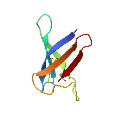
    Synthetic macrocycles such as calixarenes and cucurbiturils are increasingly applied as mediators of protein assembly and crystallization. The macrocycle can facilitate assembly by providing a surface on which two or more proteins bind simultaneously. This work explores the capacity of the sulfonato-calix[ n ]arene (sclx n ) series to effect crystallization of PAF, a small, cationic antifungal protein. Co-crystallization with sclx 4 , sclx 6 or sclx 8 led to high-resolution crystal structures. In the absence of sclx n , diffraction-quality crystals of PAF were not obtained. Interestingly, all three sclx n were bound to a similar patch on PAF. The largest and most flexible variant, sclx 8 , yielded a dimer of PAF. Complex formation was evident in solution via NMR and ITC experiments, showing more pronounced effects with increasing macrocycle size. In agreement with the crystal structure, the ITC data suggested that sclx 8 acts as a bidentate ligand. The contributions of calixarene size/conformation to protein recognition and assembly are discussed. Finally, it is suggested that the conserved binding site for anionic calixarenes implicates this region of PAF in membrane binding, which is a prerequisite for antifungal activity.


  • Organizational Affiliation

    School of Chemistry, National University of Ireland, University Road, Galway, Ireland.


Macromolecules
Find similar proteins by:  (by identity cutoff)  |  3D Structure
Entity ID: 1
MoleculeChains Sequence LengthOrganismDetailsImage
Pc24g00380 protein
A, B
55Penicillium rubens Wisconsin 54-1255Mutation(s): 0 
UniProt
Find proteins for B6HWK0 (Penicillium rubens (strain ATCC 28089 / DSM 1075 / NRRL 1951 / Wisconsin 54-1255))
Explore B6HWK0 
Go to UniProtKB:  B6HWK0
Entity Groups  
Sequence Clusters30% Identity50% Identity70% Identity90% Identity95% Identity100% Identity
UniProt GroupB6HWK0
Sequence Annotations
Expand
  • Reference Sequence
Small Molecules
Ligands 2 Unique
IDChains Name / Formula / InChI Key2D Diagram3D Interactions
EVB (Subject of Investigation/LOI)
Query on EVB

Download Ideal Coordinates CCD File 
D [auth B]sulfonato-calix[8]arene
C56 H48 O32 S8
KCEGJGDGMRAJEP-UHFFFAOYSA-N
7PE (Subject of Investigation/LOI)
Query on 7PE

Download Ideal Coordinates CCD File 
C [auth A]2-(2-(2-(2-(2-(2-ETHOXYETHOXY)ETHOXY)ETHOXY)ETHOXY)ETHOXY)ETHANOL
C14 H30 O7
UKXKPKBTMYNOFS-UHFFFAOYSA-N
Binding Affinity Annotations 
IDSourceBinding Affinity
EVB Binding MOAD:  6HAJ Kd: 1.06e+4 (nM) from 1 assay(s)
Experimental Data & Validation

Experimental Data

  • Method: X-RAY DIFFRACTION
  • Resolution: 1.50 Å
  • R-Value Free: 0.244 
  • R-Value Work: 0.217 
  • R-Value Observed: 0.220 
  • Space Group: P 61
Unit Cell:
Length ( Å )Angle ( ˚ )
a = 24.3α = 90
b = 24.3β = 90
c = 313.69γ = 120
Software Package:
Software NamePurpose
BUSTERrefinement
MOSFLMdata reduction
Aimlessdata scaling
PHASERphasing

Structure Validation

View Full Validation Report


Ligand Structure Quality Assessment 


Entry History & Funding Information

Deposition Data


Funding OrganizationLocationGrant Number
Science Foundation IrelandIreland13/CDA/2168
Science Foundation IrelandIreland13/ERC/B2912

Revision History  (Full details and data files)

  • Version 1.0: 2019-02-13
    Type: Initial release
  • Version 1.1: 2019-03-27
    Changes: Data collection, Database references
  • Version 2.0: 2019-04-17
    Changes: Atomic model, Author supporting evidence, Data collection, Derived calculations, Non-polymer description, Structure summary
  • Version 2.1: 2024-01-17
    Changes: Data collection, Database references, Refinement description