6H8P | pdb_00006h8p

JMJD2A/ KDM4A COMPLEXED WITH NI(II), NOG AND Histone H1.4(18-32)K26me3 peptide (15-mer)


Experimental Data Snapshot

  • Method: X-RAY DIFFRACTION
  • Resolution: 1.98 Å
  • R-Value Free: 
    0.212 (Depositor), 0.212 (DCC) 
  • R-Value Work: 
    0.192 (Depositor), 0.194 (DCC) 
  • R-Value Observed: 
    0.193 (Depositor) 

Starting Model: experimental
View more details

wwPDB Validation 3D Report Full Report

Validation slider image for 6H8P

This is version 1.3 of the entry. See complete history

Literature

Mechanistic and structural studies of KDM-catalysed demethylation of histone 1 isotype 4 at lysine 26.

Walport, L.J.Hopkinson, R.J.Chowdhury, R.Zhang, Y.Bonnici, J.Schiller, R.Kawamura, A.Schofield, C.J.

(2018) FEBS Lett 592: 3264-3273

  • DOI: https://doi.org/10.1002/1873-3468.13231
  • Primary Citation Related Structures: 
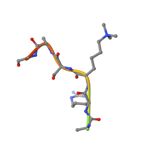
    6H8P

  • PubMed Abstract: 

    N-Methylation of lysyl residues is widely observed on histone proteins. Using isolated enzymes, we report mechanistic and structural studies on histone lysine demethylase (KDM)-catalysed demethylation of N ε -methylated lysine 26 on histone 1 isotype 4 (H1.4). The results reveal that methylated H1.4K26 is a substrate for all members of the KDM4 subfamily and that KDM4A-catalysed demethylation of H1.4K26me3 peptide is similarly efficient to that of H3K9me3. Crystallographic studies of an H1.4K26me3:KDM4A complex reveal a conserved binding geometry to that of H3K9me3. In the light of the high activity of the KDM4s on this mark, our results suggest JmjC KDM-catalysed demethylation of H1.4K26 may be as prevalent as demethylation on the H3 tail and warrants further investigation in cells.


  • Organizational Affiliation
    • Department of Chemistry, Chemistry Research Laboratory, University of Oxford, UK.

Macromolecule Content 

  • Total Structure Weight: 92.81 kDa 
  • Atom Count: 6,238 
  • Modeled Residue Count: 710 
  • Deposited Residue Count: 792 
  • Unique protein chains: 2

Macromolecules

Find similar proteins by:|  3D Structure
Entity ID: 1
MoleculeChains  Sequence LengthOrganismDetailsImage
Lysine-specific demethylase 4A
A, B
381Homo sapiensMutation(s): 0 
Gene Names: KDM4AJHDM3AJMJD2JMJD2AKIAA0677
EC: 1.14.11 (PDB Primary Data), 1.14.11.66 (UniProt), 1.14.11.69 (UniProt)
UniProt & NIH Common Fund Data Resources
Find proteins for O75164 (Homo sapiens)
Explore O75164 
Go to UniProtKB:  O75164
PHAROS:  O75164
GTEx:  ENSG00000066135 
Entity Groups
Sequence Clusters30% Identity50% Identity70% Identity90% Identity95% Identity100% Identity
UniProt GroupO75164
Sequence Annotations
Expand
Reference Sequence
Find similar proteins by:  Sequence   |   3D Structure  
Entity ID: 2
MoleculeChains  Sequence LengthOrganismDetailsImage
Histone H1.4
C, D
15Homo sapiensMutation(s): 0 
UniProt & NIH Common Fund Data Resources
Find proteins for P10412 (Homo sapiens)
Explore P10412 
Go to UniProtKB:  P10412
PHAROS:  P10412
GTEx:  ENSG00000168298 
Entity Groups
Sequence Clusters30% Identity50% Identity70% Identity90% Identity95% Identity100% Identity
UniProt GroupP10412
Sequence Annotations
Expand
Reference Sequence

Small Molecules

Ligands 5 Unique
IDChains Name / Formula / InChI Key2D Diagram3D Interactions
OGA

Query on OGA


Download:Ideal Coordinates CCD File
H [auth A],
O [auth B]
N-OXALYLGLYCINE
C4 H5 N O5
BIMZLRFONYSTPT-UHFFFAOYSA-N
GOL

Query on GOL


Download:Ideal Coordinates CCD File
I [auth A],
J [auth A],
K [auth A],
P [auth B]
GLYCEROL
C3 H8 O3
PEDCQBHIVMGVHV-UHFFFAOYSA-N
ZN

Query on ZN


Download:Ideal Coordinates CCD File
F [auth A],
M [auth B]
ZINC ION
Zn
PTFCDOFLOPIGGS-UHFFFAOYSA-N
NI

Query on NI


Download:Ideal Coordinates CCD File
E [auth A],
L [auth B]
NICKEL (II) ION
Ni
VEQPNABPJHWNSG-UHFFFAOYSA-N
CL

Query on CL


Download:Ideal Coordinates CCD File
G [auth A],
N [auth B]
CHLORIDE ION
Cl
VEXZGXHMUGYJMC-UHFFFAOYSA-M
Modified Residues  1 Unique
IDChains TypeFormula2D DiagramParent
M3L
Query on M3L
C, D
L-PEPTIDE LINKINGC9 H21 N2 O2LYS
Binding Affinity Annotations 
IDSourceBinding Affinity
OGA BindingDB:  6H8P IC50: min: 8.50e+4, max: 2.50e+5 (nM) from 2 assay(s)

Experimental Data & Validation

Experimental Data

  • Method: X-RAY DIFFRACTION
  • Resolution: 1.98 Å
  • R-Value Free:  0.212 (Depositor), 0.212 (DCC) 
  • R-Value Work:  0.192 (Depositor), 0.194 (DCC) 
  • R-Value Observed: 0.193 (Depositor) 
Space Group: P 21 21 2
Unit Cell:
Length ( Å )Angle ( ˚ )
a = 100.722α = 90
b = 150.146β = 90
c = 57.601γ = 90
Software Package:
Software NamePurpose
PHENIXrefinement
HKL-2000data reduction
HKL-2000data scaling
PHASERphasing

Structure Validation

View Full Validation Report


Entry History 

Deposition Data

Revision History  (Full details and data files)

  • Version 1.0: 2018-08-15
    Type: Initial release
  • Version 1.1: 2018-09-12
    Changes: Data collection, Database references
  • Version 1.2: 2018-10-24
    Changes: Data collection, Database references
  • Version 1.3: 2024-01-17
    Changes: Data collection, Database references, Derived calculations, Refinement description