Machining protein microcrystals for structure determination by electron diffraction.
Duyvesteyn, H.M.E., Kotecha, A., Ginn, H.M., Hecksel, C.W., Beale, E.V., de Haas, F., Evans, G., Zhang, P., Chiu, W., Stuart, D.I.(2018) Proc Natl Acad Sci U S A 115: 9569-9573
- PubMed: 30171169 Search on PubMedSearch on PubMed Central
- DOI: https://doi.org/10.1073/pnas.1809978115
- Primary Citation Related Structures:
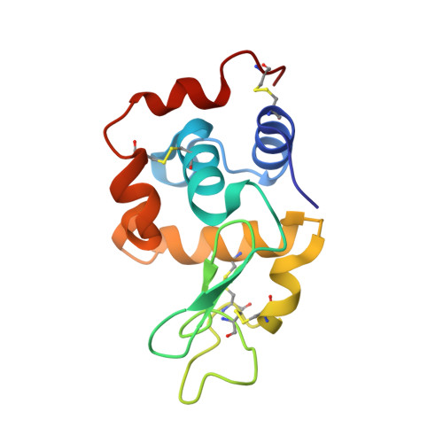
6H3B - PubMed Abstract:
We demonstrate that ion-beam milling of frozen, hydrated protein crystals to thin lamella preserves the crystal lattice to near-atomic resolution. This provides a vehicle for protein structure determination, bridging the crystal size gap between the nanometer scale of conventional electron diffraction and micron scale of synchrotron microfocus beamlines. The demonstration that atomic information can be retained suggests that milling could provide such detail on sections cut from vitrified cells.
- Division of Structural Biology, University of Oxford, Headington, Oxford OX3 7BN, United Kingdom.
Organizational Affiliation: