6H1P

Cold-adapted beta-D-galactosidase from Arthrobacter sp. 32cB - data collected at room temperature


Experimental Data Snapshot

  • Method: X-RAY DIFFRACTION
  • Resolution: 3.01 Å
  • R-Value Free: 0.229 
  • R-Value Work: 0.181 
  • R-Value Observed: 0.184 

wwPDB Validation   3D Report Full Report


This is version 1.2 of the entry. See complete history


Literature

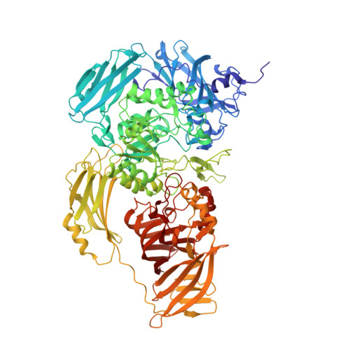
Structural features of cold-adapted dimeric GH2 beta-D-galactosidase from Arthrobacter sp. 32cB.

Rutkiewicz, M.Bujacz, A.Bujacz, G.

(2019) Biochim Biophys Acta Proteins Proteom 1867: 776-786

  • DOI: https://doi.org/10.1016/j.bbapap.2019.06.001
  • Primary Citation of Related Structures:  
    6H1P

  • PubMed Abstract: 

    Crystal structures of cold-adapted β-d-galactosidase (EC 3.2.1.23) from the Antarctic bacterium Arthrobacter sp. 32cB (ArthβDG) have been determined in an unliganded form resulting from diffraction experiments conducted at 100 K (at resolution 1.8 Å) and at room temperature (at resolution 3.0 Å). A detailed comparison of those two structures of the same enzyme was performed in order to estimate differences in their molecular flexibility and rigidity and to study structural rationalization for the cold-adaptation of the investigated enzyme. Furthermore, a comparative analysis with structures of homologous enzymes from psychrophilic, mesophilic, and thermophilic sources has been discussed to elucidate the relationship between structure and cold-adaptation in a wider context. The performed studies confirm that the structure of cold-adapted ArthβDG maintains balance between molecular stability and structural flexibility, which can be observed independently on the temperature of conducted X-ray diffraction experiments. Obtained information about proper protein function under given conditions provide a guideline for rational engineering of proteins in terms of their temperature optimum and thermal stability.


  • Organizational Affiliation

    Institute of Technical Biochemistry, Faculty of Biotechnology and Food Sciences, Lodz University of Technology, Stefanowskiego 4/10, 90-924 Lodz, Poland.


Macromolecules
Find similar proteins by:  (by identity cutoff)  |  3D Structure
Entity ID: 1
MoleculeChains Sequence LengthOrganismDetailsImage
Beta-galactosidase989Arthrobacter sp. 32cBMutation(s): 0 
EC: 3.2.1.23
UniProt
Find proteins for A0A023UGN9 (Arthrobacter sp. 32cB)
Explore A0A023UGN9 
Go to UniProtKB:  A0A023UGN9
Entity Groups  
Sequence Clusters30% Identity50% Identity70% Identity90% Identity95% Identity100% Identity
UniProt GroupA0A023UGN9
Sequence Annotations
Expand
  • Reference Sequence
Small Molecules
Ligands 1 Unique
IDChains Name / Formula / InChI Key2D Diagram3D Interactions
NA
Query on NA

Download Ideal Coordinates CCD File 
B [auth A]SODIUM ION
Na
FKNQFGJONOIPTF-UHFFFAOYSA-N
Experimental Data & Validation

Experimental Data

  • Method: X-RAY DIFFRACTION
  • Resolution: 3.01 Å
  • R-Value Free: 0.229 
  • R-Value Work: 0.181 
  • R-Value Observed: 0.184 
  • Space Group: P 31 2 1
Unit Cell:
Length ( Å )Angle ( ˚ )
a = 140.219α = 90
b = 140.219β = 90
c = 127.995γ = 120
Software Package:
Software NamePurpose
XDSdata reduction
PHENIXrefinement
PDB_EXTRACTdata extraction
Aimlessdata scaling
PHASERphasing

Structure Validation

View Full Validation Report


Entry History & Funding Information

Deposition Data


Funding OrganizationLocationGrant Number
PolandNational Science Center Poland 2016/21/B/ST5/00555

Revision History  (Full details and data files)

  • Version 1.0: 2019-06-26
    Type: Initial release
  • Version 1.1: 2019-07-24
    Changes: Data collection, Database references
  • Version 1.2: 2024-01-17
    Changes: Data collection, Database references, Refinement description