Characterization of the pleiotropic LysR-type transcription regulator LeuO of Escherichia coli.
Fragel, S.M., Montada, A., Heermann, R., Baumann, U., Schacherl, M., Schnetz, K.(2019) Nucleic Acids Res 47: 7363-7379
- PubMed: 31184713 Search on PubMedSearch on PubMed Central
- DOI: https://doi.org/10.1093/nar/gkz506
- Primary Citation Related Structures:
6GZ0, 6GZ1, 6GZ2 - PubMed Abstract:
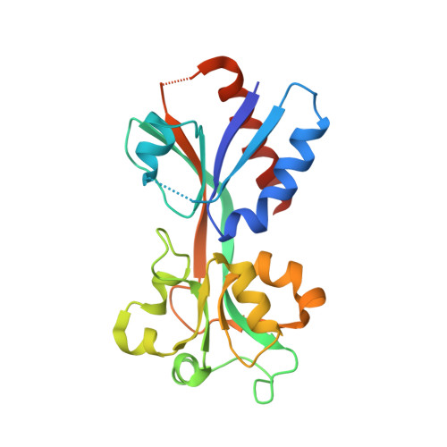
LeuO is a pleiotropic LysR-type transcriptional regulator (LTTR) and co-regulator of the abundant nucleoid-associated repressor protein H-NS in Gammaproteobacteria. As other LTTRs, LeuO is a tetramer that is formed by dimerization of the N-terminal DNA-binding domain (DBD) and C-terminal effector-binding domain (EBD). To characterize the Escherichia coli LeuO protein, we screened for LeuO mutants that activate the cas (CRISPR-associated/Cascade) promoter more effectively than wild-type LeuO. This yielded nine mutants carrying amino acid substitutions in the dimerization interface of the regulatory EBD, as shown by solving the EBD's crystal structure. Superimposing of the crystal structures of LeuO-EBD and LeuO-S120D-EBD suggests that the Ser120 to Asp substitution triggers a structural change that is related to effector-induced structural changes of LTTRs. Corresponding functional analyses demonstrated that LeuO-S120D has a higher DNA-binding affinity than wild-type LeuO. Further, a palindromic DNA-binding core-site and a consensus sequence were identified by DNase I footprinting with LeuO-S120D as well as with the dimeric DBD. The data suggest that LeuO-S120D mimics an effector-induced form of LeuO regulating a distinct set of target loci. In general, constitutive mutants and determining the DNA-binding specificity of the DBD-dimer are feasible approaches to characterize LTTRs of unknown function.
- Institute for Genetics, University of Cologne, Zülpicher Str. 47a, 50674 Cologne, Germany.
Organizational Affiliation: