6GVE

GAPDH-CP12-PRK complex


Experimental Data Snapshot

  • Method: ELECTRON MICROSCOPY
  • Resolution: 3.90 Å
  • Aggregation State: PARTICLE 
  • Reconstruction Method: SINGLE PARTICLE 

wwPDB Validation   3D Report Full Report


This is version 1.2 of the entry. See complete history


Literature

Structural basis of light-induced redox regulation in the Calvin-Benson cycle in cyanobacteria.

McFarlane, C.R.Shah, N.R.Kabasakal, B.V.Echeverria, B.Cotton, C.A.R.Bubeck, D.Murray, J.W.

(2019) Proc Natl Acad Sci U S A 116: 20984-20990

  • DOI: https://doi.org/10.1073/pnas.1906722116
  • Primary Citation of Related Structures:  
    6GFO, 6GFP, 6GFQ, 6GFR, 6GG7, 6GHL, 6GHR, 6GVE

  • PubMed Abstract: 

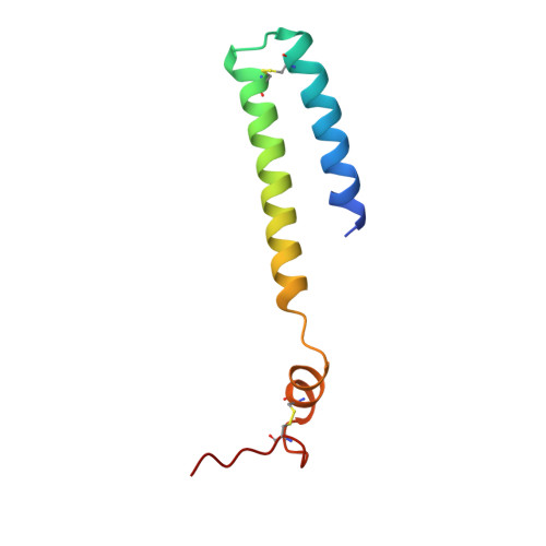
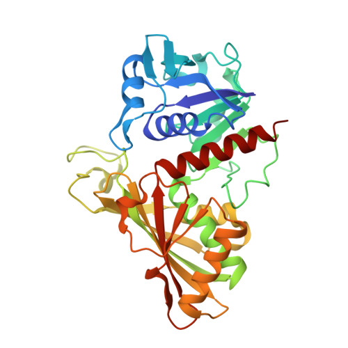
    Plants, algae, and cyanobacteria fix carbon dioxide to organic carbon with the Calvin-Benson (CB) cycle. Phosphoribulokinase (PRK) and glyceraldehyde 3-phosphate dehydrogenase (GAPDH) are essential CB-cycle enzymes that control substrate availability for the carboxylation enzyme Rubisco. PRK consumes ATP to produce the Rubisco substrate ribulose bisphosphate (RuBP). GAPDH catalyzes the reduction step of the CB cycle with NADPH to produce the sugar glyceraldehyde 3-phosphate (GAP), which is used for regeneration of RuBP and is the main exit point of the cycle. GAPDH and PRK are coregulated by the redox state of a conditionally disordered protein CP12, which forms a ternary complex with both enzymes. However, the structural basis of CB-cycle regulation by CP12 is unknown. Here, we show how CP12 modulates the activity of both GAPDH and PRK. Using thermophilic cyanobacterial homologs, we solve crystal structures of GAPDH with different cofactors and CP12 bound, and the ternary GAPDH-CP12-PRK complex by electron cryo-microscopy, we reveal that formation of the N-terminal disulfide preorders CP12 prior to binding the PRK active site, which is resolved in complex with CP12. We find that CP12 binding to GAPDH influences substrate accessibility of all GAPDH active sites in the binary and ternary inhibited complexes. Our structural and biochemical data explain how CP12 integrates responses from both redox state and nicotinamide dinucleotide availability to regulate carbon fixation.


  • Organizational Affiliation

    Department of Life Sciences, Imperial College London, SW7 2AZ, United Kingdom.


Macromolecules
Find similar proteins by:  (by identity cutoff)  |  3D Structure
Entity ID: 1
MoleculeChains Sequence LengthOrganismDetailsImage
CP12 polypeptideA [auth P],
N [auth C],
O [auth H],
P [auth M]
77Thermosynechococcus vestitus BP-1Mutation(s): 0 
Gene Names: cp12
UniProt
Find proteins for Q8DHX3 (Thermosynechococcus vestitus (strain NIES-2133 / IAM M-273 / BP-1))
Explore Q8DHX3 
Go to UniProtKB:  Q8DHX3
Entity Groups  
Sequence Clusters30% Identity50% Identity70% Identity90% Identity95% Identity100% Identity
UniProt GroupQ8DHX3
Sequence Annotations
Expand
  • Reference Sequence
Find similar proteins by:  (by identity cutoff)  |  3D Structure
Entity ID: 2
MoleculeChains Sequence LengthOrganismDetailsImage
Glyceraldehyde-3-phosphate dehydrogenase339Thermosynechococcus vestitus BP-1Mutation(s): 0 
Gene Names: tll1466
EC: 1.2.1
UniProt
Find proteins for Q8DIW5 (Thermosynechococcus vestitus (strain NIES-2133 / IAM M-273 / BP-1))
Explore Q8DIW5 
Go to UniProtKB:  Q8DIW5
Entity Groups  
Sequence Clusters30% Identity50% Identity70% Identity90% Identity95% Identity100% Identity
UniProt GroupQ8DIW5
Sequence Annotations
Expand
  • Reference Sequence
Find similar proteins by:  (by identity cutoff)  |  3D Structure
Entity ID: 3
MoleculeChains Sequence LengthOrganismDetailsImage
PhosphoribulokinaseJ,
K [auth B],
L [auth G],
M [auth L]
334Thermosynechococcus vestitus BP-1Mutation(s): 0 
EC: 2.7.1.19
UniProt
Find proteins for Q8DHN2 (Thermosynechococcus vestitus (strain NIES-2133 / IAM M-273 / BP-1))
Explore Q8DHN2 
Go to UniProtKB:  Q8DHN2
Entity Groups  
Sequence Clusters30% Identity50% Identity70% Identity90% Identity95% Identity100% Identity
UniProt GroupQ8DHN2
Sequence Annotations
Expand
  • Reference Sequence
Experimental Data & Validation

Experimental Data

  • Method: ELECTRON MICROSCOPY
  • Resolution: 3.90 Å
  • Aggregation State: PARTICLE 
  • Reconstruction Method: SINGLE PARTICLE 
EM Software:
TaskSoftware PackageVersion
MODEL REFINEMENTPHENIX1.13

Structure Validation

View Full Validation Report


Entry History & Funding Information

Deposition Data


Funding OrganizationLocationGrant Number
Biotechnology and Biological Sciences Research CouncilUnited KingdomBB/J014575/1

Revision History  (Full details and data files)

  • Version 1.0: 2019-07-03
    Type: Initial release
  • Version 1.1: 2019-11-20
    Changes: Data collection, Database references
  • Version 1.2: 2019-12-11
    Changes: Other