Biochemical properties of a Pseudomonas aminotransferase involved in caprolactam metabolism.
Palacio, C.M., Rozeboom, H.J., Lanfranchi, E., Meng, Q., Otzen, M., Janssen, D.B.(2019) FEBS J 286: 4086-4102
- PubMed: 31162815 Search on PubMed
- DOI: https://doi.org/10.1111/febs.14950
- Primary Citation Related Structures:
6G4B, 6G4C, 6G4D, 6G4E, 6G4F - PubMed Abstract:
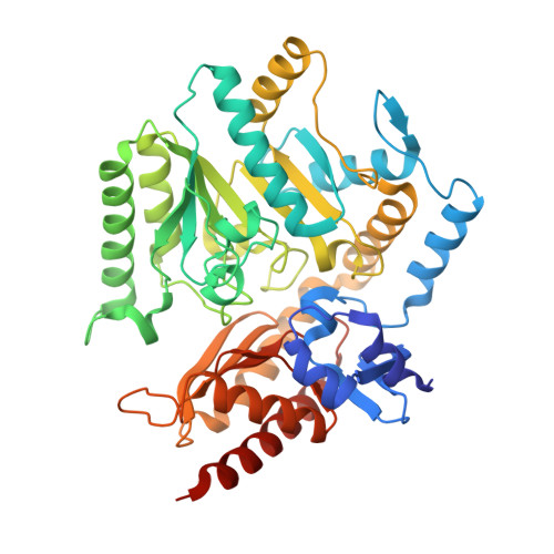
The biodegradation of the nylon-6 precursor caprolactam by a strain of Pseudomonas jessenii proceeds via ATP-dependent hydrolytic ring opening to 6-aminohexanoate. This non-natural ω-amino acid is converted to 6-oxohexanoic acid by an aminotransferase (PjAT) belonging to the fold type I pyridoxal 5'-phosphate (PLP) enzymes. To understand the structural basis of 6-aminohexanoatate conversion, we solved different crystal structures and determined the substrate scope with a range of aliphatic and aromatic amines. Comparison with the homologous aminotransferases from Chromobacterium violaceum (CvAT) and Vibrio fluvialis (VfAT) showed that the PjAT enzyme has the lowest K M values (highest affinity) and highest specificity constant (k cat /K M ) with the caprolactam degradation intermediates 6-aminohexanoate and 6-oxohexanoic acid, in accordance with its proposed in vivo function. Five distinct three-dimensional structures of PjAT were solved by protein crystallography. The structure of the aldimine intermediate formed from 6-aminohexanoate and the PLP cofactor revealed the presence of a narrow hydrophobic substrate-binding tunnel leading to the cofactor and covered by a flexible arginine, which explains the high activity and selectivity of the PjAT with 6-aminohexanoate. The results suggest that the degradation pathway for caprolactam has recruited an aminotransferase that is well adapted to 6-aminohexanoate degradation. DATABASE: The atomic coordinates and structure factors P. jessenii 6-aminohexanoate aminotransferase have been deposited in the PDB as entries 6G4B (E∙succinate complex), 6G4C (E∙phosphate complex), 6G4D (E∙PLP complex), 6G4E (E∙PLP-6-aminohexanoate intermediate), and 6G4F (E∙PMP complex).
- Biotransformation and Biocatalysis, Groningen Biomolecular Sciences and Biotechnology Institute (GBB), University of Groningen, The Netherlands.
Organizational Affiliation: