6G2T | pdb_00006g2t

human cardiac myosin binding protein C C1 Ig-domain bound to native cardiac thin filament


Experimental Data Snapshot

  • Method: ELECTRON MICROSCOPY
  • Resolution: 9.00 Å
  • Aggregation State: HELICAL ARRAY 
  • Reconstruction Method: HELICAL 

wwPDB Validation 3D Report Full Report

Validation slider image for 6G2T

This is version 1.2 of the entry. See complete history

Literature

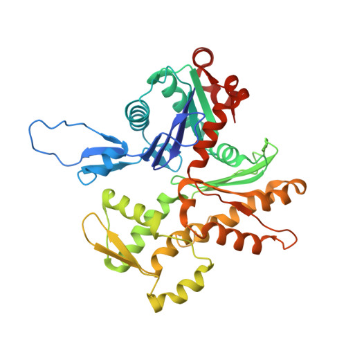
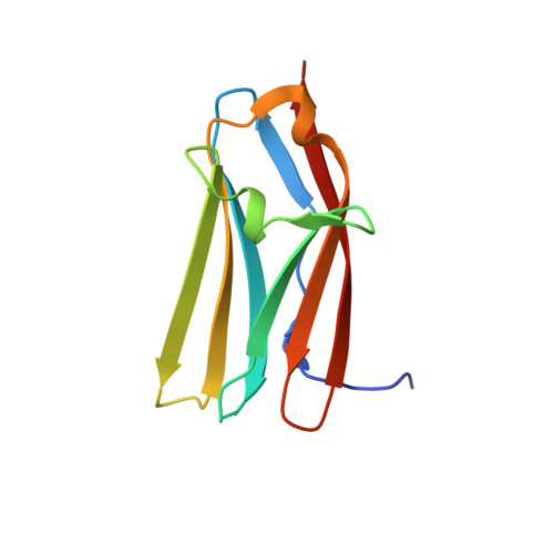
N-Terminal Domains of Cardiac Myosin Binding Protein C Cooperatively Activate the Thin Filament.

Risi, C.Belknap, B.Forgacs-Lonart, E.Harris, S.P.Schroder, G.F.White, H.D.Galkin, V.E.

(2018) Structure 26: 1604-1611.e4

  • DOI: https://doi.org/10.1016/j.str.2018.08.007
  • Primary Citation Related Structures: 
    6CXI, 6CXJ, 6G2T

  • PubMed Abstract: 

    Muscle contraction relies on interaction between myosin-based thick filaments and actin-based thin filaments. Myosin binding protein C (MyBP-C) is a key regulator of actomyosin interactions. Recent studies established that the N'-terminal domains (NTDs) of MyBP-C can either activate or inhibit thin filaments, but the mechanism of their collective action is poorly understood. Cardiac MyBP-C (cMyBP-C) harbors an extra NTD, which is absent in skeletal isoforms of MyBP-C, and its role in regulation of cardiac contraction is unknown. Here we show that the first two domains of human cMyPB-C (i.e., C0 and C1) cooperate to activate the thin filament. We demonstrate that C1 interacts with tropomyosin via a positively charged loop and that this interaction, stabilized by the C0 domain, is required for thin filament activation by cMyBP-C. Our data reveal a mechanism by which cMyBP-C can modulate cardiac contraction and demonstrate a function of the C0 domain.


  • Organizational Affiliation
    • Department of Physiological Sciences, Eastern Virginia Medical School, 700 West Olney Road, Lewis Hall, Room 3126, Norfolk, VA 23507, USA.

Macromolecule Content 

  • Total Structure Weight: 370.15 kDa 
  • Atom Count: 24,958 
  • Modeled Residue Count: 3,384 
  • Deposited Residue Count: 3,444 
  • Unique protein chains: 3

Macromolecules

Find similar proteins by:|  3D Structure
Entity ID: 1
MoleculeChains  Sequence LengthOrganismDetailsImage
Actin, cytoplasmic 2
A, B, C, D, E
A, B, C, D, E, F
375Homo sapiensMutation(s): 0 
Gene Names: ACTG1ACTG
EC: 3.6.4
UniProt & NIH Common Fund Data Resources
Find proteins for P63261 (Homo sapiens)
Explore P63261 
Go to UniProtKB:  P63261
PHAROS:  P63261
GTEx:  ENSG00000184009 
Entity Groups
Sequence Clusters30% Identity50% Identity70% Identity90% Identity95% Identity100% Identity
UniProt GroupP63261
Sequence Annotations
Expand
Reference Sequence
Find similar proteins by:|  3D Structure
Entity ID: 2
MoleculeChains  Sequence LengthOrganismDetailsImage
Myosin-binding protein C, cardiac-type
G, H, I, J, K
G, H, I, J, K, L
109Homo sapiensMutation(s): 0 
Gene Names: MYBPC3
UniProt & NIH Common Fund Data Resources
Find proteins for Q14896 (Homo sapiens)
Explore Q14896 
Go to UniProtKB:  Q14896
PHAROS:  Q14896
GTEx:  ENSG00000134571 
Entity Groups
Sequence Clusters30% Identity50% Identity70% Identity90% Identity95% Identity100% Identity
UniProt GroupQ14896
Sequence Annotations
Expand
Reference Sequence
Find similar proteins by:|  3D Structure
Entity ID: 3
MoleculeChains  Sequence LengthOrganismDetailsImage
Tropomyosin
M, N, O, P
135Sus scrofaMutation(s): 0 

Experimental Data & Validation

Experimental Data

  • Method: ELECTRON MICROSCOPY
  • Resolution: 9.00 Å
  • Aggregation State: HELICAL ARRAY 
  • Reconstruction Method: HELICAL 
EM Software:
TaskSoftware PackageVersion
RECONSTRUCTIONSPIDER
MODEL REFINEMENTCNS

Structure Validation

View Full Validation Report


Entry History 

& Funding Information

Deposition Data


Funding OrganizationLocationGrant Number
American Heart AssociationUnited States16GRNT31220040

Revision History  (Full details and data files)

  • Version 1.0: 2018-10-17
    Type: Initial release
  • Version 1.1: 2018-12-19
    Changes: Data collection, Database references, Structure summary
  • Version 1.2: 2024-05-15
    Changes: Data collection, Database references