Structure-Based Optimization Strategies for G Protein-Coupled Receptor (GPCR) Allosteric Modulators: A Case Study from Analyses of New Metabotropic Glutamate Receptor 5 (mGlu5) X-ray Structures.
Christopher, J.A., Orgovan, Z., Congreve, M., Dore, A.S., Errey, J.C., Marshall, F.H., Mason, J.S., Okrasa, K., Rucktooa, P., Serrano-Vega, M.J., Ferenczy, G.G., Keseru, G.M.(2019) J Med Chem 62: 207-222
- PubMed: 29455526 Search on PubMed
- DOI: https://doi.org/10.1021/acs.jmedchem.7b01722
- Primary Citation Related Structures:
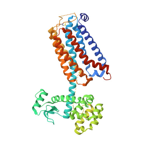
6FFH, 6FFI - PubMed Abstract:
Two interesting new X-ray structures of negative allosteric modulator (NAM) ligands for the mGlu 5 receptor, M-MPEP (3) and fenobam (4), are reported. The new structures show how the binding of the ligands induces different receptor water channel conformations to previously published structures. The structure of fenobam, where a urea replaces the acetylenic linker in M-MPEP and mavoglurant, reveals a binding mode where the ligand is rotated by 180° compared to a previously proposed docking model. The need for multiple ligand structures for accurate GPCR structure-based drug design is demonstrated by the different growing vectors identified for the head groups of M-MPEP and mavoglurant and by the unexpected water-mediated receptor interactions of a new chemotype represented by fenobam. The implications of the new structures for ligand design are discussed, with extensive analysis of the energetics of the water networks of both pseudoapo and bound structures providing a new design strategy for allosteric modulators.
- Heptares Therapeutics Ltd. , BioPark , Welwyn Garden City , Hertfordshire AL7 3AX , U.K.
Organizational Affiliation: