6F7Y

Crystal structure of dimethylated RSL, cucurbituril-free form


Experimental Data Snapshot

  • Method: X-RAY DIFFRACTION
  • Resolution: 1.60 Å
  • R-Value Free: 0.199 
  • R-Value Work: 0.169 
  • R-Value Observed: 0.170 

wwPDB Validation   3D Report Full Report


This is version 2.1 of the entry. See complete history


Literature

Cucurbit[7]uril-Dimethyllysine Recognition in a Model Protein.

Guagnini, F.Antonik, P.M.Rennie, M.L.O'Byrne, P.Khan, A.R.Pinalli, R.Dalcanale, E.Crowley, P.B.

(2018) Angew Chem Int Ed Engl 57: 7126-7130

  • DOI: https://doi.org/10.1002/anie.201803232
  • Primary Citation of Related Structures:  
    6F7W, 6F7X, 6F7Y

  • PubMed Abstract: 

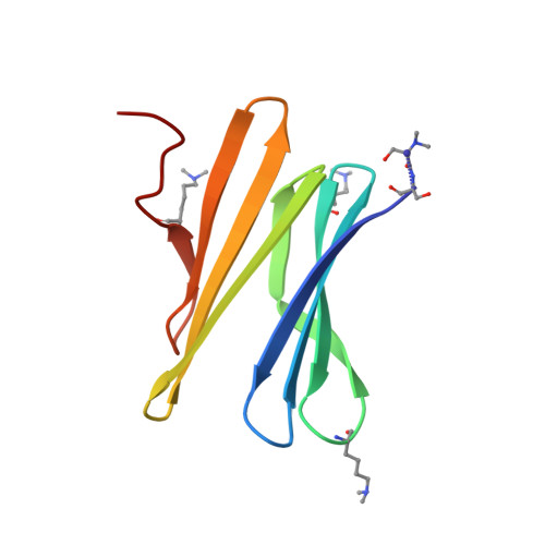
    Here, we provide the first structural characterization of host-guest complexation between cucurbit[7]uril (Q7) and dimethyllysine (KMe 2 ) in a model protein. Binding was dominated by complete encapsulation of the dimethylammonium functional group. While selectivity for the most sterically accessible dimethyllysine was observed both in solution and in the solid state, three different modes of Q7-KMe 2 complexation were revealed by X-ray crystallography. The crystal structures revealed also entrapped water molecules that solvated the ammonium group within the Q7 cavity. Remarkable Q7-protein assemblies, including inter-locked octahedral cages that comprise 24 protein trimers, occurred in the solid state. Cucurbituril clusters appear to be responsible for these assemblies, suggesting a strategy to generate controlled protein architectures.


  • Organizational Affiliation

    School of Chemistry, National University of Ireland Galway, University Road, Galway, Ireland.


Macromolecules
Find similar proteins by:  (by identity cutoff)  |  3D Structure
Entity ID: 1
MoleculeChains Sequence LengthOrganismDetailsImage
Fucose-binding lectin protein
A, B, C, D
90Ralstonia solanacearumMutation(s): 0 
Gene Names: RSP795_21825RSP799_05830RUN39_v1_50103
UniProt
Find proteins for A0A0S4TLR1 (Ralstonia solanacearum)
Explore A0A0S4TLR1 
Go to UniProtKB:  A0A0S4TLR1
Entity Groups  
Sequence Clusters30% Identity50% Identity70% Identity90% Identity95% Identity100% Identity
UniProt GroupA0A0S4TLR1
Sequence Annotations
Expand
  • Reference Sequence
Experimental Data & Validation

Experimental Data

  • Method: X-RAY DIFFRACTION
  • Resolution: 1.60 Å
  • R-Value Free: 0.199 
  • R-Value Work: 0.169 
  • R-Value Observed: 0.170 
  • Space Group: P 63
Unit Cell:
Length ( Å )Angle ( ˚ )
a = 122.285α = 90
b = 122.285β = 90
c = 36.418γ = 120
Software Package:
Software NamePurpose
XDSdata reduction
XSCALEdata scaling
PHASERphasing
REFMACrefinement
PDB_EXTRACTdata extraction

Structure Validation

View Full Validation Report


Entry History & Funding Information

Deposition Data


Funding OrganizationLocationGrant Number
Science Foundation IrelandIreland13/ERC/B2912 and 13/CDA/2168

Revision History  (Full details and data files)

  • Version 1.0: 2018-05-30
    Type: Initial release
  • Version 1.1: 2018-06-13
    Changes: Data collection, Database references
  • Version 2.0: 2020-08-05
    Changes: Data collection, Polymer sequence
  • Version 2.1: 2024-01-17
    Changes: Data collection, Database references, Refinement description