6F3F | pdb_00006f3f

Autoinhibited Src kinase bound to ADP


Experimental Data Snapshot

  • Method: X-RAY DIFFRACTION
  • Resolution: 2.42 Å
  • R-Value Free: 
    0.268 (Depositor), 0.268 (DCC) 
  • R-Value Work: 
    0.208 (Depositor), 0.208 (DCC) 
  • R-Value Observed: 
    0.211 (Depositor) 

Starting Model: experimental
View more details

wwPDB Validation 3D Report Full Report

Validation slider image for 6F3F

Ligand Structure Quality Assessment 


This is version 1.2 of the entry. See complete history

Literature

A switch in nucleotide affinity governs activation of the Src and Tec family kinases.

von Raussendorf, F.de Ruiter, A.Leonard, T.A.

(2017) Sci Rep 7: 17405-17405

  • DOI: https://doi.org/10.1038/s41598-017-17703-5
  • Primary Citation Related Structures: 
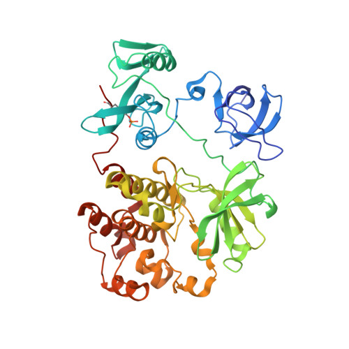
    6F3F

  • PubMed Abstract: 

    The Tec kinases, closely related to Src family kinases, are essential for lymphocyte function in the adaptive immune system. Whilst the Src and Abl kinases are regulated by tail phosphorylation and N-terminal myristoylation respectively, the Tec kinases are notable for the absence of either regulatory element. We have found that the inactive conformations of the Tec kinase Itk and Src preferentially bind ADP over ATP, stabilising both proteins. We demonstrate that Itk adopts the same conformation as Src and that the autoinhibited conformation of Src is independent of its C-terminal tail. Allosteric activation of both Itk and Src depends critically on the disruption of a conserved hydrophobic stack that accompanies regulatory domain displacement. We show that a conformational switch permits the exchange of ADP for ATP, leading to efficient autophosphorylation and full activation. In summary, we propose a universal mechanism for the activation and autoinhibition of the Src and Tec kinases.


  • Organizational Affiliation
    • Department of Structural and Computational Biology, Max F. Perutz Laboratories (MFPL), Campus Vienna Biocenter 5, 1030, Vienna, Austria.

Macromolecule Content 

  • Total Structure Weight: 52.49 kDa 
  • Atom Count: 3,729 
  • Modeled Residue Count: 449 
  • Deposited Residue Count: 455 
  • Unique protein chains: 1

Macromolecules

Find similar proteins by:|  3D Structure
Entity ID: 1
MoleculeChains  Sequence LengthOrganismDetailsImage
Tyrosine-protein kinase455Mus musculusMutation(s): 0 
Gene Names: Src
EC: 2.7.10.2
UniProt & NIH Common Fund Data Resources
Find proteins for P05480 (Mus musculus)
Explore P05480 
Go to UniProtKB:  P05480
IMPC:  MGI:98397
Entity Groups
Sequence Clusters30% Identity50% Identity70% Identity90% Identity95% Identity100% Identity
UniProt GroupP05480
Sequence Annotations
Expand
Reference Sequence

Small Molecules

Modified Residues  1 Unique
IDChains TypeFormula2D DiagramParent
PTR
Query on PTR
A
L-PEPTIDE LINKINGC9 H12 N O6 PTYR

Experimental Data & Validation

Experimental Data

  • Method: X-RAY DIFFRACTION
  • Resolution: 2.42 Å
  • R-Value Free:  0.268 (Depositor), 0.268 (DCC) 
  • R-Value Work:  0.208 (Depositor), 0.208 (DCC) 
  • R-Value Observed: 0.211 (Depositor) 
Space Group: P 21 21 21
Unit Cell:
Length ( Å )Angle ( ˚ )
a = 51.011α = 90
b = 82.974β = 90
c = 105.051γ = 90
Software Package:
Software NamePurpose
PHENIXrefinement
MOSFLMdata reduction
Aimlessdata scaling
PHASERphasing

Structure Validation

View Full Validation Report


Ligand Structure Quality Assessment 


Entry History 

& Funding Information

Deposition Data


Funding OrganizationLocationGrant Number
Austrian Science FundAustriaP28135

Revision History  (Full details and data files)

  • Version 1.0: 2017-12-27
    Type: Initial release
  • Version 1.1: 2024-01-17
    Changes: Data collection, Database references, Derived calculations, Refinement description
  • Version 1.2: 2024-10-23
    Changes: Structure summary