6ENY

Structure of the human PLC editing module


Experimental Data Snapshot

  • Method: ELECTRON MICROSCOPY
  • Resolution: 5.80 Å
  • Aggregation State: PARTICLE 
  • Reconstruction Method: SINGLE PARTICLE 

wwPDB Validation   3D Report Full Report


This is version 2.0 of the entry. See complete history


Literature

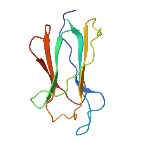
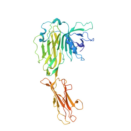
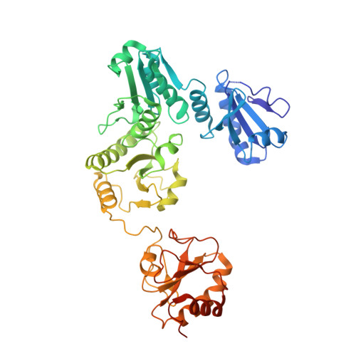
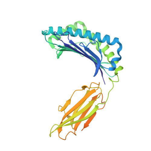
Structure of the human MHC-I peptide-loading complex.

Blees, A.Januliene, D.Hofmann, T.Koller, N.Schmidt, C.Trowitzsch, S.Moeller, A.Tampe, R.

(2017) Nature 551: 525-528

  • DOI: https://doi.org/10.1038/nature24627
  • Primary Citation of Related Structures:  
    6ENY

  • PubMed Abstract: 

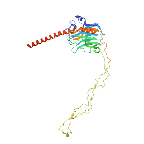
    The peptide-loading complex (PLC) is a transient, multisubunit membrane complex in the endoplasmic reticulum that is essential for establishing a hierarchical immune response. The PLC coordinates peptide translocation into the endoplasmic reticulum with loading and editing of major histocompatibility complex class I (MHC-I) molecules. After final proofreading in the PLC, stable peptide-MHC-I complexes are released to the cell surface to evoke a T-cell response against infected or malignant cells. Sampling of different MHC-I allomorphs requires the precise coordination of seven different subunits in a single macromolecular assembly, including the transporter associated with antigen processing (TAP1 and TAP2, jointly referred to as TAP), the oxidoreductase ERp57, the MHC-I heterodimer, and the chaperones tapasin and calreticulin. The molecular organization of and mechanistic events that take place in the PLC are unknown owing to the heterogeneous composition and intrinsically dynamic nature of the complex. Here, we isolate human PLC from Burkitt's lymphoma cells using an engineered viral inhibitor as bait and determine the structure of native PLC by electron cryo-microscopy. Two endoplasmic reticulum-resident editing modules composed of tapasin, calreticulin, ERp57, and MHC-I are centred around TAP in a pseudo-symmetric orientation. A multivalent chaperone network within and across the editing modules establishes the proofreading function at two lateral binding platforms for MHC-I molecules. The lectin-like domain of calreticulin senses the MHC-I glycan, whereas the P domain reaches over the MHC-I peptide-binding pocket towards ERp57. This arrangement allows tapasin to facilitate peptide editing by clamping MHC-I. The translocation pathway of TAP opens out into a large endoplasmic reticulum lumenal cavity, confined by the membrane entry points of tapasin and MHC-I. Two lateral windows channel the antigenic peptides to MHC-I. Structures of PLC captured at distinct assembly states provide mechanistic insight into the recruitment and release of MHC-I. Our work defines the molecular symbiosis of an ABC transporter and an endoplasmic reticulum chaperone network in MHC-I assembly and provides insight into the onset of the adaptive immune response.


  • Organizational Affiliation

    Institute of Biochemistry, Biocenter, Goethe University Frankfurt, Max-von-Laue Strasse 9, 60438 Frankfurt/Main, Germany.


Macromolecules
Find similar proteins by:  (by identity cutoff)  |  3D Structure
Entity ID: 1
MoleculeChains Sequence LengthOrganismDetailsImage
Beta-2-microglobulinA [auth B]99Homo sapiensMutation(s): 0 
UniProt & NIH Common Fund Data Resources
Find proteins for P61769 (Homo sapiens)
Explore P61769 
Go to UniProtKB:  P61769
PHAROS:  P61769
GTEx:  ENSG00000166710 
Entity Groups  
Sequence Clusters30% Identity50% Identity70% Identity90% Identity95% Identity100% Identity
UniProt GroupP61769
Sequence Annotations
Expand
  • Reference Sequence
Find similar proteins by:  (by identity cutoff)  |  3D Structure
Entity ID: 2
MoleculeChains Sequence LengthOrganismDetailsImage
TapasinB [auth C]428Homo sapiensMutation(s): 0 
UniProt & NIH Common Fund Data Resources
Find proteins for O15533 (Homo sapiens)
Explore O15533 
Go to UniProtKB:  O15533
PHAROS:  O15533
GTEx:  ENSG00000231925 
Entity Groups  
Sequence Clusters30% Identity50% Identity70% Identity90% Identity95% Identity100% Identity
UniProt GroupO15533
Sequence Annotations
Expand
  • Reference Sequence
Find similar proteins by:  (by identity cutoff)  |  3D Structure
Entity ID: 3
MoleculeChains Sequence LengthOrganismDetailsImage
Protein disulfide-isomerase A3C [auth D]481Homo sapiensMutation(s): 0 
EC: 5.3.4.1
UniProt & NIH Common Fund Data Resources
Find proteins for P30101 (Homo sapiens)
Explore P30101 
Go to UniProtKB:  P30101
PHAROS:  P30101
GTEx:  ENSG00000167004 
Entity Groups  
Sequence Clusters30% Identity50% Identity70% Identity90% Identity95% Identity100% Identity
UniProt GroupP30101
Sequence Annotations
Expand
  • Reference Sequence
Find similar proteins by:  (by identity cutoff)  |  3D Structure
Entity ID: 4
MoleculeChains Sequence LengthOrganismDetailsImage
HLA class I histocompatibility antigen, A-3 alpha chainD [auth F]341Homo sapiensMutation(s): 0 
UniProt & NIH Common Fund Data Resources
Find proteins for P04439 (Homo sapiens)
Explore P04439 
Go to UniProtKB:  P04439
PHAROS:  P04439
GTEx:  ENSG00000206503 
Entity Groups  
Sequence Clusters30% Identity50% Identity70% Identity90% Identity95% Identity100% Identity
UniProt GroupP04439
Sequence Annotations
Expand
  • Reference Sequence
Find similar proteins by:  (by identity cutoff)  |  3D Structure
Entity ID: 5
MoleculeChains Sequence LengthOrganismDetailsImage
CalreticulinE [auth G]400Homo sapiensMutation(s): 0 
UniProt & NIH Common Fund Data Resources
Find proteins for P27797 (Homo sapiens)
Explore P27797 
Go to UniProtKB:  P27797
PHAROS:  P27797
GTEx:  ENSG00000179218 
Entity Groups  
Sequence Clusters30% Identity50% Identity70% Identity90% Identity95% Identity100% Identity
UniProt GroupP27797
Sequence Annotations
Expand
  • Reference Sequence
Oligosaccharides

Help

Entity ID: 6
MoleculeChains Length2D Diagram Glycosylation3D Interactions
2-acetamido-2-deoxy-beta-D-glucopyranose-(1-4)-2-acetamido-2-deoxy-beta-D-glucopyranoseF [auth A]2N/A
Glycosylation Resources
GlyTouCan:  G42666HT
GlyCosmos:  G42666HT
GlyGen:  G42666HT
Entity ID: 7
MoleculeChains Length2D Diagram Glycosylation3D Interactions
beta-D-glucopyranose-(1-3)-alpha-D-mannopyranose-(1-2)-alpha-D-mannopyranose-(1-2)-alpha-D-mannopyranoseG [auth E]4N/AN/A
Experimental Data & Validation

Experimental Data

  • Method: ELECTRON MICROSCOPY
  • Resolution: 5.80 Å
  • Aggregation State: PARTICLE 
  • Reconstruction Method: SINGLE PARTICLE 
EM Software:
TaskSoftware PackageVersion
RECONSTRUCTIONFREALIGNX
MODEL REFINEMENTPHENIX

Structure Validation

View Full Validation Report


Entry History & Funding Information

Deposition Data


Funding OrganizationLocationGrant Number
German Research FoundationGermanySFB 807
German Research FoundationGermanyGRK 1986

Revision History  (Full details and data files)

  • Version 1.0: 2017-11-29
    Type: Initial release
  • Version 1.1: 2017-12-06
    Changes: Database references
  • Version 2.0: 2020-07-29
    Type: Remediation
    Reason: Carbohydrate remediation
    Changes: Advisory, Atomic model, Data collection, Derived calculations, Structure summary