Structure-Function Characterization of Streptococcus intermedius Surface Antigen Pas.
Mieher, J.L., Schormann, N., Wu, R., Patel, M., Purushotham, S., Wu, H., Scoffield, J., Deivanayagam, C.(2021) J Bacteriol 203: e0017521-e0017521
- PubMed: 34339301 Search on PubMedSearch on PubMed Central
- DOI: https://doi.org/10.1128/JB.00175-21
- Primary Citation Related Structures:
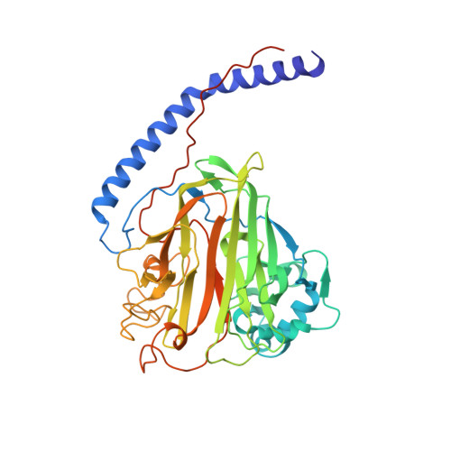
6E36, 6E3F - PubMed Abstract:
Streptococcus intermedius, an oral commensal bacterium, is found at various sites, including subgingival dental plaque, purulent infections, and cystic fibrosis lungs. Oral streptococci utilize proteins on their surface to adhere to tissues and/or surfaces localizing the bacteria, which subsequently leads to the development of biofilms, colonization, and infection. Among the 19 genomically annotated cell wall-attached surface proteins on S. intermedius, Pas is an adhesin that belongs to the antigen I/II (AgI/II) family. Here, we have structurally and functionally characterized Pas, particularly focusing on its microbial-host as well as microbial-microbial interactions. The crystal structures of V Pas and C 123 Pas show high similarity with AgI/II of Streptococcus mutans. V Pas hosts a conserved metal binding site, and likewise, the C 123 Pas structure retains its conserved metal binding sites and isopeptide bonds within its three DEv-IgG domains. Pas interacts with nanomolar affinity to lung alveolar glycoprotein 340 (Gp340), its scavenger receptor cysteine-rich domains (SRCRs), and with fibrinogen. Both Candida albicans and Pseudomonas aeruginosa, the opportunistic pathogens that cohabitate with S. intermedius in the lungs of CFTR patients were studied in dual-species biofilm studies. The Pas-deficient mutant (Δ pas ) displayed significant reduction in dual-biofilm formation with C. albicans. In similar studies with P. aeruginosa, Pas did not mediate the biofilm formation with either the acute isolate (PAO1) or the chronic isolate (FRD1). However, the sortase A-deficient mutant (Δ srtA ) displayed reduced biofilm formation with both C. albicans and P. aeruginosa FRD1. Taken together, our findings highlight the role of Pas in both microbial-host and interkingdom interactions and expose its potential role in disease outcomes. IMPORTANCE Streptococcus intermedius, an oral commensal bacterium, has been clinically observed in subgingival dental plaque, purulent infections, and cystic fibrosis lungs. In this study, we have (i) determined the crystal structure of the V and C regions of Pas; (ii) shown that its surface protein Pas adheres to fibrinogen, which could potentially ferry the microbe through the bloodstream from the oral cavity; (iii) characterized Pas's high-affinity adherence to lung alveolar protein Gp340 that could fixate the microbe on lung epithelial cells; and (iv) most importantly, shown that these surface proteins on the oral commensal S. intermedius enhance biofilms of known pathogens Candida albicans and Pseudomonas aeruginosa.
- Department of Biochemistry and Molecular Genetics, University of Alabama at Birminghamgrid.265892.2, Birmingham, Alabama, USA.
Organizational Affiliation: