6E00

Structure of a N-Me-p-iodo-D-Phe1,N-Me-D-Gln4,Lys10-teixobactin analogue

  • Classification: ANTIBIOTIC
  • Organism(s): Eleftheria terrae
  • Mutation(s): No 

  • Deposited: 2018-07-05 Released: 2018-10-17 
  • Deposition Author(s): Nowick, J.S., Yang, H., Wierzbicki, M.
  • Funding Organization(s): National Institutes of Health/National Institute Of Allergy and Infectious Diseases (NIH/NIAID)

Experimental Data Snapshot

  • Method: X-RAY DIFFRACTION
  • Resolution: 2.20 Å
  • R-Value Free: 0.246 
  • R-Value Work: 0.211 
  • R-Value Observed: 0.213 

wwPDB Validation   3D Report Full Report


This is version 2.0 of the entry. See complete history


Literature

X-ray Crystallographic Structure of a Teixobactin Derivative Reveals Amyloid-like Assembly.

Yang, H.Wierzbicki, M.Du Bois, D.R.Nowick, J.S.

(2018) J Am Chem Soc 140: 14028-14032

  • DOI: https://doi.org/10.1021/jacs.8b07709
  • Primary Citation of Related Structures:  
    6E00

  • PubMed Abstract: 

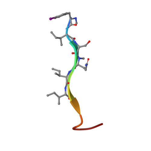
    This paper describes the X-ray crystallographic structure of a derivative of the antibiotic teixobactin and shows that its supramolecular assembly through the formation of antiparallel β-sheets creates binding sites for oxyanions. An active derivative of teixobactin containing lysine in place of allo-enduracididine assembles to form amyloid-like fibrils, which are observed through a thioflavin T fluorescence assay and by transmission electron microscopy. A homologue, bearing an N-methyl substituent, to attenuate fibril formation, and an iodine atom, to facilitate X-ray crystallographic phase determination, crystallizes as double helices of β-sheets that bind sulfate anions. β-Sheet dimers are key subunits of these assemblies, with the N-terminal methylammonium group of one monomer and the C-terminal macrocycle of the other monomer binding each anion. These observations suggest a working model for the mechanism of action of teixobactin, in which the antibiotic assembles and the assemblies bind lipid II and related bacterial cell wall precursors on the surface of Gram-positive bacteria.


  • Organizational Affiliation

    Department of Chemistry , University of California, Irvine , Irvine , California 92697-2025 , United States.


Macromolecules

Find similar proteins by:  Sequence   |   3D Structure  

Entity ID: 1
MoleculeChains Sequence LengthOrganismDetailsImage
N-Me-p-iodo-D-Phe1,N-Me-D-Gln4,Lys10-teixobactin analogue11Eleftheria terraeMutation(s): 0 
Entity Groups  
Sequence Clusters30% Identity50% Identity70% Identity90% Identity95% Identity100% Identity
Sequence Annotations
Expand
  • Reference Sequence
Small Molecules
Ligands 1 Unique
IDChains Name / Formula / InChI Key2D Diagram3D Interactions
SO4
Query on SO4

Download Ideal Coordinates CCD File 
AB [auth e]
BB [auth f]
CB [auth g]
DB [auth h]
EB [auth i]
AB [auth e],
BB [auth f],
CB [auth g],
DB [auth h],
EB [auth i],
FB [auth j],
GA [auth A],
GB [auth k],
HA [auth B],
HB [auth l],
IA [auth C],
IB [auth m],
JA [auth D],
JB [auth n],
KA [auth E],
KB [auth o],
LA [auth F],
LB [auth p],
MA [auth G],
NA [auth H],
OA [auth I],
PA [auth J],
QA [auth K],
RA [auth L],
SA [auth M],
TA [auth N],
UA [auth O],
VA [auth P],
WA [auth a],
XA [auth b],
YA [auth c],
ZA [auth d]
SULFATE ION
O4 S
QAOWNCQODCNURD-UHFFFAOYSA-L
Biologically Interesting Molecules (External Reference) 1 Unique
Entity ID: 1
IDChains NameType/Class2D Diagram3D Interactions
PRD_002317
Query on PRD_002317
A
AA [auth B]
B [auth C]
BA [auth D]
C [auth E]
A,
AA [auth B],
B [auth C],
BA [auth D],
C [auth E],
CA [auth F],
D [auth G],
DA [auth H],
E [auth I],
EA [auth J],
F [auth K],
FA [auth L],
G [auth M],
H [auth N],
I [auth O],
J [auth P],
K [auth a],
L [auth b],
M [auth c],
N [auth d],
O [auth e],
P [auth f],
Q [auth g],
R [auth h],
S [auth i],
T [auth j],
U [auth k],
V [auth l],
W [auth m],
X [auth n],
Y [auth o],
Z [auth p]
N-Me-p-iodo-D-Phe1,N-Me-D-Gln4,Lys10-teixobactin analoguePeptide-like / Antibiotic
Experimental Data & Validation

Experimental Data

  • Method: X-RAY DIFFRACTION
  • Resolution: 2.20 Å
  • R-Value Free: 0.246 
  • R-Value Work: 0.211 
  • R-Value Observed: 0.213 
  • Space Group: P 21 21 21
Unit Cell:
Length ( Å )Angle ( ˚ )
a = 47.478α = 90
b = 69.37β = 90
c = 115.422γ = 90
Software Package:
Software NamePurpose
REFMACrefinement
XDSdata reduction
Aimlessdata scaling
PHENIXphasing

Structure Validation

View Full Validation Report


Entry History & Funding Information

Deposition Data


Funding OrganizationLocationGrant Number
National Institutes of Health/National Institute Of Allergy and Infectious Diseases (NIH/NIAID)United States1R21AI121548-01

Revision History  (Full details and data files)

  • Version 1.0: 2018-10-17
    Type: Initial release
  • Version 1.1: 2018-11-14
    Changes: Data collection, Database references
  • Version 1.2: 2019-12-18
    Changes: Author supporting evidence
  • Version 2.0: 2023-11-15
    Changes: Atomic model, Data collection, Database references, Derived calculations