6DN1

CRYSTAL STRUCTURE OF THE FMN RIBOSWITCH BOUND TO BRX1151 SPLIT RNA


Experimental Data Snapshot

  • Method: X-RAY DIFFRACTION
  • Resolution: 3.03 Å
  • R-Value Free: 0.242 
  • R-Value Work: 0.223 
  • R-Value Observed: 0.224 

wwPDB Validation   3D Report Full Report


Ligand Structure Quality Assessment 


This is version 1.4 of the entry. See complete history


Literature

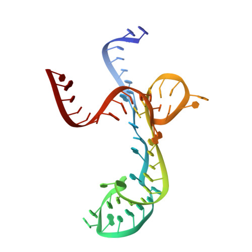
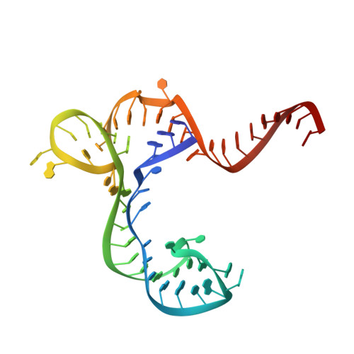
Structure-Activity Relationship of Flavin Analogues That Target the Flavin Mononucleotide Riboswitch.

Vicens, Q.Mondragon, E.Reyes, F.E.Coish, P.Aristoff, P.Berman, J.Kaur, H.Kells, K.W.Wickens, P.Wilson, J.Gadwood, R.C.Schostarez, H.J.Suto, R.K.Blount, K.F.Batey, R.T.

(2018) ACS Chem Biol 13: 2908-2919

  • DOI: https://doi.org/10.1021/acschembio.8b00533
  • Primary Citation of Related Structures:  
    6DN1, 6DN2, 6DN3

  • PubMed Abstract: 

    The flavin mononucleotide (FMN) riboswitch is an emerging target for the development of novel RNA-targeting antibiotics. We previously discovered an FMN derivative, 5FDQD, that protects mice against diarrhea-causing Clostridium difficile bacteria. Here, we present the structure-based drug design strategy that led to the discovery of this fluoro-phenyl derivative with antibacterial properties. This approach involved the following stages: (1) structural analysis of all available free and bound FMN riboswitch structures; (2) design, synthesis, and purification of derivatives; (3) in vitro testing for productive binding using two chemical probing methods; (4) in vitro transcription termination assays; and (5) resolution of the crystal structures of the FMN riboswitch in complex with the most mature candidates. In the process, we delineated principles for productive binding to this riboswitch, thereby demonstrating the effectiveness of a coordinated structure-guided approach to designing drugs against RNA.


  • Organizational Affiliation

    Department of Chemistry and Biochemistry , University of Colorado , 596 UCB , Boulder , Colorado 80309 , United States.


Macromolecules
Find similar nucleic acids by:  (by identity cutoff)  |  3D Structure
Entity ID: 1
MoleculeChains LengthOrganismImage
RNA RIBOSWITCHA [auth X]54Fusobacterium nucleatum
Sequence Annotations
Expand
  • Reference Sequence
Find similar nucleic acids by:  (by identity cutoff)  |  3D Structure
Entity ID: 2
MoleculeChains LengthOrganismImage
RNA (57-MER)B [auth Z]57Fusobacterium nucleatum
Sequence Annotations
Expand
  • Reference Sequence
Small Molecules
Ligands 3 Unique
IDChains Name / Formula / InChI Key2D Diagram3D Interactions
GZ7 (Subject of Investigation/LOI)
Query on GZ7

Download Ideal Coordinates CCD File 
G [auth Z]10-(6-carboxyhexyl)-8-(cyclopentylamino)-2,4-dihydroxy-7-methylbenzo[g]pteridin-10-ium
C23 H30 N5 O4
AJTKEKBUVMMJJR-UHFFFAOYSA-O
K
Query on K

Download Ideal Coordinates CCD File 
H [auth Z]POTASSIUM ION
K
NPYPAHLBTDXSSS-UHFFFAOYSA-N
MG
Query on MG

Download Ideal Coordinates CCD File 
C [auth X]
D [auth X]
E [auth X]
F [auth X]
I [auth Z]
C [auth X],
D [auth X],
E [auth X],
F [auth X],
I [auth Z],
J [auth Z]
MAGNESIUM ION
Mg
JLVVSXFLKOJNIY-UHFFFAOYSA-N
Experimental Data & Validation

Experimental Data

  • Method: X-RAY DIFFRACTION
  • Resolution: 3.03 Å
  • R-Value Free: 0.242 
  • R-Value Work: 0.223 
  • R-Value Observed: 0.224 
  • Space Group: P 31 2 1
Unit Cell:
Length ( Å )Angle ( ˚ )
a = 74.44α = 90
b = 74.44β = 90
c = 140.71γ = 120
Software Package:
Software NamePurpose
XSCALEdata scaling
BUSTERrefinement
PDB_EXTRACTdata extraction
XDSdata reduction
PHASERphasing

Structure Validation

View Full Validation Report


Ligand Structure Quality Assessment 


Entry History 

Revision History  (Full details and data files)

  • Version 1.0: 2018-09-05
    Type: Initial release
  • Version 1.1: 2018-10-03
    Changes: Data collection, Database references
  • Version 1.2: 2018-10-31
    Changes: Data collection, Database references
  • Version 1.3: 2020-10-21
    Changes: Data collection
  • Version 1.4: 2023-10-11
    Changes: Data collection, Database references, Refinement description