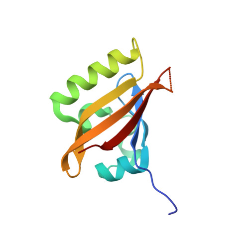
Structural basis of DSF recognition by its receptor RpfR and its regulatory interaction with the DSF synthase RpfF.
Waldron, E.J., Snyder, D., Fernandez, N.L., Sileo, E., Inoyama, D., Freundlich, J.S., Waters, C.M., Cooper, V.S., Neiditch, M.B.(2019) PLoS Biol 17: e3000123-e3000123
- PubMed: 30716063 Search on PubMedSearch on PubMed Central
- DOI: https://doi.org/10.1371/journal.pbio.3000123
- Primary Citation Related Structures:
6DGA, 6DGG, 6DGJ, 6DGN - PubMed Abstract:
The diffusible signal factors (DSFs) are a family of quorum-sensing autoinducers (AIs) produced and detected by numerous gram-negative bacteria. The DSF family AIs are fatty acids, differing in their acyl chain length, branching, and substitution but having in common a cis-2 double bond that is required for their activity. In both human and plant pathogens, DSFs regulate diverse phenotypes, including virulence factor expression, antibiotic resistance, and biofilm dispersal. Despite their widespread relevance to both human health and agriculture, the molecular basis of DSF recognition by their cellular receptors remained a mystery. Here, we report the first structure-function studies of the DSF receptor regulation of pathogenicity factor R (RpfR). We present the X-ray crystal structure of the RpfR DSF-binding domain in complex with the Burkholderia DSF (BDSF), which to our knowledge is the first structure of a DSF receptor in complex with its AI. To begin to understand the mechanistic role of the BDSF-RpfR contacts observed in the biologically important complex, we have also determined the X-ray crystal structure of the RpfR DSF-binding domain in complex with the inactive, saturated isomer of BDSF, dodecanoic acid (C12:0). In addition to these ligand-receptor complex structures, we report the discovery of a previously overlooked RpfR domain and show that it binds to and negatively regulates the DSF synthase regulation of pathogenicity factor F (RpfF). We have named this RpfR region the RpfF interaction (FI) domain, and we have determined its X-ray crystal structure alone and in complex with RpfF. These X-ray crystal structures, together with extensive complementary in vivo and in vitro functional studies, reveal the molecular basis of DSF recognition and the importance of the cis-2 double bond to DSF function. Finally, we show that throughout cellular growth, the production of BDSF by RpfF is post-translationally controlled by the RpfR N-terminal FI domain, affecting the cellular concentration of the bacterial second messenger bis-(3'-5')-cyclic dimeric guanosine monophosphate (c-di-GMP). Thus, in addition to describing the molecular basis for the binding and specificity of a DSF for its receptor, we describe a receptor-synthase interaction regulating bacterial quorum-sensing signaling and second messenger signal transduction.
- Department of Microbiology, Biochemistry, and Molecular Genetics, New Jersey Medical School, Rutgers, State University of New Jersey, Newark, New Jersey, United States of America.
Organizational Affiliation: