6DF1

Anti-phosphotyrosine antibody 4G10-4D5 Fab complexed with phosphotyrosine peptide


Experimental Data Snapshot

  • Method: X-RAY DIFFRACTION
  • Resolution: 2.30 Å
  • R-Value Free: 0.222 
  • R-Value Work: 0.203 
  • R-Value Observed: 0.204 

wwPDB Validation   3D Report Full Report


This is version 1.3 of the entry. See complete history


Literature

Engineering Improved Antiphosphotyrosine Antibodies Based on an Immunoconvergent Binding Motif.

Mou, Y.Zhou, X.X.Leung, K.Martinko, A.J.Yu, J.Y.Chen, W.Wells, J.A.

(2018) J Am Chem Soc 140: 16615-16624

  • DOI: https://doi.org/10.1021/jacs.8b08402
  • Primary Citation of Related Structures:  
    6DEZ, 6DF0, 6DF1, 6DF2

  • PubMed Abstract: 

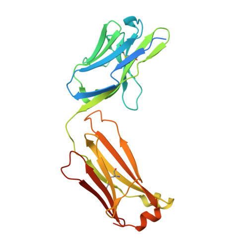
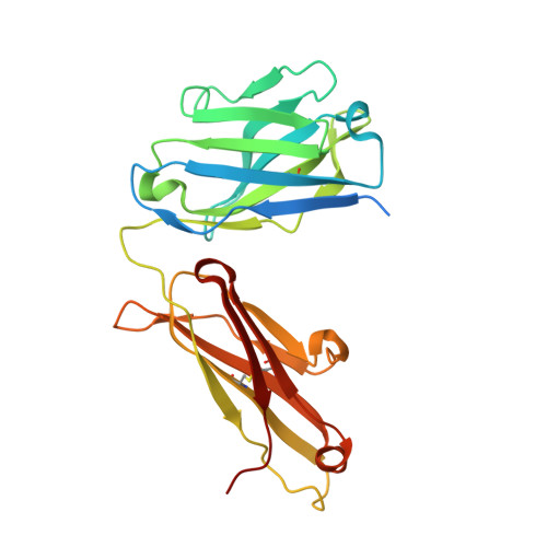
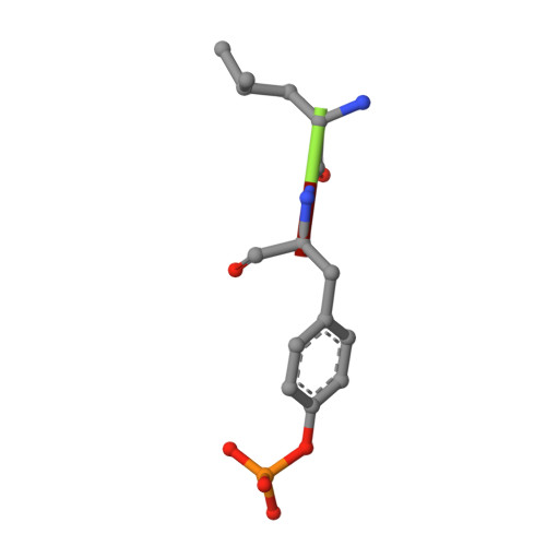
    Phosphotyrosine (pY) is one of the most highly studied posttranslational modifications that is responsible for tightly regulating many signaling pathways in eukaryotes. Pan-specific pY antibodies have emerged as powerful tools for understanding the role of these modifications. Nevertheless, structures have not been reported for pan-specific pY antibodies, greatly impeding the further development of tools for integrating this ubiquitous posttranslational modification using structure-guided designs. Here, we present the first crystal structures of two widely utilized pan-specific pY antibodies, PY20 and 4G10. The two antibodies, although developed independently from animal immunizations, have surprisingly similar modes of recognition of the phosphate group, implicating a generic binding structure among pan-specific pY antibodies. Sequence alignments revealed that many pY binding residues are predominant in the mouse V germline genes, which consequently led to the convergent antibodies. On the basis of the convergent structure, we designed a phage display library by lengthening the CDR-L3 loop with the aid of computational modeling. Panning with this library resulted in a series of 4G10 variants with 4 to 11-fold improvements in pY binding affinities. The crystal structure of one improved variant showed remarkable superposition to the computational model, where the lengthened CDR-L3 loop creates an additional hydrogen bond indirectly bound to the phosphate group via a water molecule. The engineered variants exhibited superior performance in Western blot and immunofluorescence.


  • Organizational Affiliation

    Department of Pharmaceutical Chemistry , University of California, San Francisco , San Francisco , California 94143 , United States.


Macromolecules
Find similar proteins by:  (by identity cutoff)  |  3D Structure
Entity ID: 1
MoleculeChains Sequence LengthOrganismDetailsImage
Anti-phosphotyrosine antibody 4G10-4D5 heavy chainA [auth L]240Homo sapiensMutation(s): 0 
Entity Groups  
Sequence Clusters30% Identity50% Identity70% Identity90% Identity95% Identity100% Identity
Sequence Annotations
Expand
  • Reference Sequence
Find similar proteins by:  (by identity cutoff)  |  3D Structure
Entity ID: 2
MoleculeChains Sequence LengthOrganismDetailsImage
Anti-phosphotyrosine antibody 4G10-4D5 light chainB [auth H]247Homo sapiensMutation(s): 0 
Entity Groups  
Sequence Clusters30% Identity50% Identity70% Identity90% Identity95% Identity100% Identity
Sequence Annotations
Expand
  • Reference Sequence

Find similar proteins by:  Sequence   |   3D Structure  

Entity ID: 3
MoleculeChains Sequence LengthOrganismDetailsImage
LEU-PTRC [auth A]3synthetic constructMutation(s): 0 
Sequence Annotations
Expand
  • Reference Sequence
Small Molecules
Modified Residues  1 Unique
IDChains TypeFormula2D DiagramParent
PTR
Query on PTR
C [auth A]L-PEPTIDE LINKINGC9 H12 N O6 PTYR
Experimental Data & Validation

Experimental Data

  • Method: X-RAY DIFFRACTION
  • Resolution: 2.30 Å
  • R-Value Free: 0.222 
  • R-Value Work: 0.203 
  • R-Value Observed: 0.204 
  • Space Group: P 21 21 21
Unit Cell:
Length ( Å )Angle ( ˚ )
a = 62.42α = 90
b = 98.93β = 90
c = 103.9γ = 90
Software Package:
Software NamePurpose
SCALAdata scaling
PHENIXrefinement
PDB_EXTRACTdata extraction
MOSFLMdata reduction
PHENIXphasing

Structure Validation

View Full Validation Report


Entry History & Funding Information

Deposition Data


Funding OrganizationLocationGrant Number
National Institutes of Health/National Cancer Institute (NIH/NCI)United StatesCA196276

Revision History  (Full details and data files)

  • Version 1.0: 2018-11-28
    Type: Initial release
  • Version 1.1: 2018-12-05
    Changes: Data collection, Structure summary
  • Version 1.2: 2019-05-01
    Changes: Data collection, Database references
  • Version 1.3: 2019-12-04
    Changes: Author supporting evidence