6D23

GLUCOSE-6-P DEHYDROGENASE (APO FORM) FROM TRYPANOSOMA CRUZI


Experimental Data Snapshot

  • Method: X-RAY DIFFRACTION
  • Resolution: 2.85 Å
  • R-Value Free: 0.249 
  • R-Value Work: 0.204 
  • R-Value Observed: 0.205 

wwPDB Validation   3D Report Full Report


This is version 1.4 of the entry. See complete history


Literature

Glucose-6-Phosphate Dehydrogenase from the Human Pathogen Trypanosoma cruzi Evolved Unique Structural Features to Support Efficient Product Formation.

Ortiz, C.Botti, H.Buschiazzo, A.Comini, M.A.

(2019) J Mol Biol 431: 2143-2162

  • DOI: https://doi.org/10.1016/j.jmb.2019.03.023
  • Primary Citation of Related Structures:  
    6D23, 6D24

  • PubMed Abstract: 

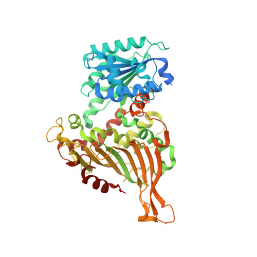
    Glucose-6-phosphate dehydrogenase (G6PDH) is the key enzyme supplying reducing power (NADPH) to the cells, by oxidation of glucose-6-phosphate (G6P), and in the process providing a precursor of ribose-5-phosphate. G6PDH is also a virulence factor of pathogenic trypanosomatid parasites. To uncover the biochemical and structural features that distinguish TcG6PDH from its human homolog, we have solved and analyzed the crystal structures of the G6PDH from Trypanosoma cruzi (TcG6PDH), alone and in complex with G6P. TcG6PDH crystallized as a tetramer and enzymatic assays further indicated that the tetramer is the active form in the parasite, in contrast to human G6PDH, which displays higher activity as a dimer. This quaternary structure was shown to be particularly stable. The molecular reasons behind this disparity were unveiled by structural analyses: a TcG6PDH-specific residue, R323, is located at the dimer-dimer interface, critically contributing with two salt bridges per subunit that are absent in the human enzyme. This explains why TcG6PDH dimerization impaired enzyme activity. The parasite protein is also distinct in displaying a 37-amino-acid extension at the N-terminus, which comprises the non-conserved C8 and C34 involved in the covalent linkage of two neighboring protomers. In addition, a cysteine triad (C53, C94 and C135) specific of Kinetoplastid G6PDHs proved critical for stabilization of TcG6PDH active site. Based on the structural and biochemical data, we posit that the N-terminal region and the catalytic site are highly dynamic. The unique structural features of TcG6PDH pave the way toward the design of efficacious and highly specific anti-trypanosomal drugs.


  • Organizational Affiliation

    Laboratory Redox Biology of Trypanosomes, Institut Pasteur de Montevideo, Mataojo 2020, 11400 Montevideo, Uruguay.


Macromolecules
Find similar proteins by:  (by identity cutoff)  |  3D Structure
Entity ID: 1
MoleculeChains Sequence LengthOrganismDetailsImage
Glucose-6-phosphate 1-dehydrogenase
A, B, C, D
541Trypanosoma cruziMutation(s): 0 
EC: 1.1.1.49
UniProt
Find proteins for Q1WBU6 (Trypanosoma cruzi)
Explore Q1WBU6 
Go to UniProtKB:  Q1WBU6
Entity Groups  
Sequence Clusters30% Identity50% Identity70% Identity90% Identity95% Identity100% Identity
UniProt GroupQ1WBU6
Sequence Annotations
Expand
  • Reference Sequence
Small Molecules
Ligands 3 Unique
IDChains Name / Formula / InChI Key2D Diagram3D Interactions
SO4
Query on SO4

Download Ideal Coordinates CCD File 
CB [auth D]
DB [auth D]
E [auth A]
EB [auth D]
F [auth A]
CB [auth D],
DB [auth D],
E [auth A],
EB [auth D],
F [auth A],
FB [auth D],
G [auth A],
GB [auth D],
H [auth A],
HB [auth D],
I [auth A],
IB [auth D],
J [auth A],
JB [auth D],
MA [auth C],
NA [auth C],
OA [auth C],
PA [auth C],
U [auth B],
V [auth B],
W [auth B],
X [auth B],
Y [auth B],
Z [auth B]
SULFATE ION
O4 S
QAOWNCQODCNURD-UHFFFAOYSA-L
GOL
Query on GOL

Download Ideal Coordinates CCD File 
AA [auth B]
BA [auth B]
CA [auth B]
HA [auth B]
IA [auth B]
AA [auth B],
BA [auth B],
CA [auth B],
HA [auth B],
IA [auth B],
JA [auth C],
K [auth A],
KA [auth C],
KB [auth D],
L [auth A],
LB [auth D],
M [auth A],
MB [auth D],
N [auth A],
NB [auth D],
O [auth A],
QA [auth C],
RA [auth C],
SA [auth C],
TA [auth C]
GLYCEROL
C3 H8 O3
PEDCQBHIVMGVHV-UHFFFAOYSA-N
CL
Query on CL

Download Ideal Coordinates CCD File 
AB [auth C]
BB [auth C]
DA [auth B]
EA [auth B]
FA [auth B]
AB [auth C],
BB [auth C],
DA [auth B],
EA [auth B],
FA [auth B],
GA [auth B],
LA [auth C],
OB [auth D],
P [auth A],
PB [auth D],
Q [auth A],
QB [auth D],
R [auth A],
RB [auth D],
S [auth A],
SB [auth D],
T [auth A],
UA [auth C],
VA [auth C],
WA [auth C],
XA [auth C],
YA [auth C],
ZA [auth C]
CHLORIDE ION
Cl
VEXZGXHMUGYJMC-UHFFFAOYSA-M
Experimental Data & Validation

Experimental Data

  • Method: X-RAY DIFFRACTION
  • Resolution: 2.85 Å
  • R-Value Free: 0.249 
  • R-Value Work: 0.204 
  • R-Value Observed: 0.205 
  • Space Group: P 1 21 1
Unit Cell:
Length ( Å )Angle ( ˚ )
a = 96.61α = 90
b = 132.96β = 100.11
c = 107.83γ = 90
Software Package:
Software NamePurpose
BUSTERrefinement
SCALAdata scaling
PDB_EXTRACTdata extraction
TRUNCATEdata reduction
AMoREphasing

Structure Validation

View Full Validation Report


Entry History 

Deposition Data

Revision History  (Full details and data files)

  • Version 1.0: 2018-05-02
    Type: Initial release
  • Version 1.1: 2018-08-15
    Changes: Data collection, Structure summary
  • Version 1.2: 2019-04-17
    Changes: Data collection, Database references
  • Version 1.3: 2019-06-05
    Changes: Data collection, Database references
  • Version 1.4: 2023-10-04
    Changes: Data collection, Database references, Refinement description