6CNU | pdb_00006cnu

Crystal Structure of JzTX-V


Experimental Data Snapshot

  • Method: X-RAY DIFFRACTION
  • Resolution: 1.05 Å
  • R-Value Free: 
    0.209 (Depositor), 0.208 (DCC) 
  • R-Value Work: 
    0.190 (Depositor), 0.190 (DCC) 
  • R-Value Observed: 
    0.191 (Depositor) 

wwPDB Validation 3D Report Full Report

Validation slider image for 6CNU

This is version 2.1 of the entry. See complete history

Literature

Discovery of Tarantula Venom-Derived NaV1.7-Inhibitory JzTx-V Peptide 5-Br-Trp24 Analogue AM-6120 with Systemic Block of Histamine-Induced Pruritis.

Wu, B.Murray, J.K.Andrews, K.L.Sham, K.Long, J.Aral, J.Ligutti, J.Amagasu, S.Liu, D.Zou, A.Min, X.Wang, Z.Ilch, C.P.Kornecook, T.J.Lin, M.J.Be, X.Miranda, L.P.Moyer, B.D.Biswas, K.

(2018) J Med Chem 61: 9500-9512

  • DOI: https://doi.org/10.1021/acs.jmedchem.8b00736
  • Primary Citation Related Structures: 
    6CNU

  • PubMed Abstract: 

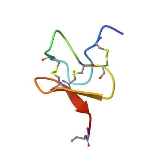
    Inhibitors of the voltage-gated sodium channel Na V 1.7 are being investigated as pain therapeutics due to compelling human genetics. We previously identified Na V 1.7-inhibitory peptides GpTx-1 and JzTx-V from tarantula venom screens. Potency and selectivity were modulated through attribute-based positional scans of native residues via chemical synthesis. Herein, we report JzTx-V lead optimization to identify a pharmacodynamically active peptide variant. Molecular docking of peptide ensembles from NMR into a homology model-derived Na V 1.7 structure supported prioritization of key residues clustered on a hydrophobic face of the disulfide-rich folded peptide for derivatization. Replacing Trp24 with 5-Br-Trp24 identified lead peptides with activity in electrophysiology assays in engineered and neuronal cells. 5-Br-Trp24 containing peptide AM-6120 was characterized in X-ray crystallography and pharmacokinetic studies and blocked histamine-induced pruritis in mice after subcutaneous administration, demonstrating systemic Na V 1.7-dependent pharmacodynamics. Our data suggests a need for high target coverage based on plasma exposure for impacting in vivo end points with selectivity-optimized peptidic Na V 1.7 inhibitors.


  • Organizational Affiliation
    • Therapeutic Discovery, Amgen Research , Amgen Inc. , 1120 Veterans Blvd , South San Francisco , California 94080 , United States.

Macromolecule Content 

  • Total Structure Weight: 7.85 kDa 
  • Atom Count: 611 
  • Modeled Residue Count: 62 
  • Deposited Residue Count: 62 
  • Unique protein chains: 2

Macromolecules

Find similar proteins by:|  3D Structure
Entity ID: 1
MoleculeChains  Sequence LengthOrganismDetailsImage
JzTx-V(D)31Chilobrachys guangxiensisMutation(s): 0 
Find similar proteins by:|  3D Structure
Entity ID: 2
MoleculeChains  Sequence LengthOrganismDetailsImage
JzTx-V31Chilobrachys guangxiensisMutation(s): 1 
Membrane Entity: Yes 
UniProt
Find proteins for Q2PAY4 (Chilobrachys guangxiensis)
Explore Q2PAY4 
Go to UniProtKB:  Q2PAY4
Entity Groups
Sequence Clusters30% Identity50% Identity70% Identity90% Identity95% Identity100% Identity
UniProt GroupQ2PAY4
Sequence Annotations
Expand
Reference Sequence

Small Molecules

Modified Residues  1 Unique
IDChains TypeFormula2D DiagramParent
DAL
Query on DAL
A
D-PEPTIDE LINKINGC3 H7 N O2

--

DAR
Query on DAR
A
D-PEPTIDE LINKINGC6 H15 N4 O2

--

DAS
Query on DAS
A
D-PEPTIDE LINKINGC4 H7 N O4

--

DCY
Query on DCY
A
D-PEPTIDE LINKINGC3 H7 N O2 S

--

DGL
Query on DGL
A
D-PEPTIDE LINKINGC5 H9 N O4

--

DGN
Query on DGN
A
D-PEPTIDE LINKINGC5 H10 N2 O3

--

DIL
Query on DIL
A
D-PEPTIDE LINKINGC6 H13 N O2

--

DLE
Query on DLE
A
D-PEPTIDE LINKINGC6 H13 N O2

--

DLY
Query on DLY
A
D-PEPTIDE LINKINGC6 H14 N2 O2

--

DSN
Query on DSN
A
D-PEPTIDE LINKINGC3 H7 N O3

--

DTH
Query on DTH
A
D-PEPTIDE LINKINGC4 H9 N O3

--

DTR
Query on DTR
A
D-PEPTIDE LINKINGC11 H12 N2 O2

--

F9D
Query on F9D
A
D-PEPTIDE LINKINGC5 H7 N O2

--

MED
Query on MED
A
D-PEPTIDE LINKINGC5 H11 N O2 S

--

LPH
Query on LPH
B
L-PEPTIDE LINKINGC5 H7 N O2

--

MSE
Query on MSE
B
L-PEPTIDE LINKINGC5 H11 N O2 SeMET

Experimental Data & Validation

Experimental Data

  • Method: X-RAY DIFFRACTION
  • Resolution: 1.05 Å
  • R-Value Free:  0.209 (Depositor), 0.208 (DCC) 
  • R-Value Work:  0.190 (Depositor), 0.190 (DCC) 
  • R-Value Observed: 0.191 (Depositor) 
Space Group: P 3 1 2
Unit Cell:
Length ( Å )Angle ( ˚ )
a = 37.16α = 90
b = 37.16β = 90
c = 64.605γ = 120
Software Package:
Software NamePurpose
REFMACrefinement
XDSdata reduction
Aimlessdata scaling
PDB_EXTRACTdata extraction
ACORNphasing

Structure Validation

View Full Validation Report


Entry History 

Deposition Data

Revision History  (Full details and data files)

  • Version 1.0: 2019-03-06
    Type: Initial release
  • Version 1.1: 2019-07-03
    Changes: Data collection, Database references, Derived calculations, Source and taxonomy
  • Version 2.0: 2020-03-04
    Type: Coordinate replacement
    Reason: Ligand identity
    Changes: Advisory, Atomic model, Data collection, Database references, Derived calculations, Non-polymer description, Polymer sequence, Refinement description, Source and taxonomy, Structure summary
  • Version 2.1: 2025-04-02
    Changes: Data collection, Database references, Derived calculations, Structure summary