6BZ5 | pdb_00006bz5

Structure and mechanism of salicylate hydroxylase from Pseudomonas putida G7


Experimental Data Snapshot

  • Method: X-RAY DIFFRACTION
  • Resolution: 2.01 Å
  • R-Value Free: 
    0.234 (Depositor), 0.266 (DCC) 
  • R-Value Work: 
    0.211 (Depositor) 
  • R-Value Observed: 
    0.212 (Depositor) 

wwPDB Validation 3D Report Full Report

Validation slider image for 6BZ5

This is version 1.3 of the entry. See complete history

Literature

Catalytic mechanism for the conversion of salicylate into catechol by the flavin-dependent monooxygenase salicylate hydroxylase.

Costa, D.M.A.Gomez, S.V.de Araujo, S.S.Pereira, M.S.Alves, R.B.Favaro, D.C.Hengge, A.C.Nagem, R.A.P.Brandao, T.A.S.

(2019) Int J Biol Macromol 129: 588-600

  • DOI: https://doi.org/10.1016/j.ijbiomac.2019.01.135
  • Primary Citation Related Structures: 
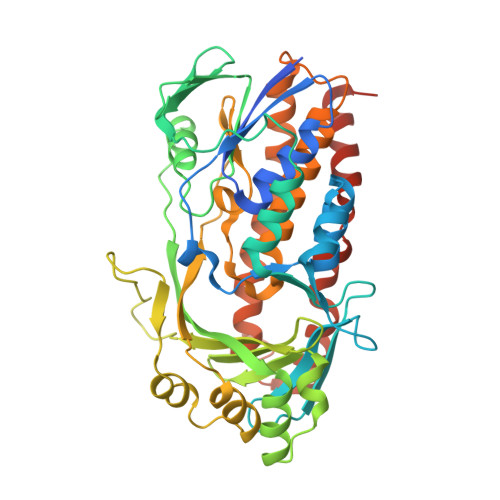
    6BZ5

  • PubMed Abstract: 

    Salicylate hydroxylase (NahG) is a flavin-dependent monooxygenase that catalyzes the decarboxylative hydroxylation of salicylate into catechol in the naphthalene degradation pathway in Pseudomonas putida G7. We explored the mechanism of action of this enzyme in detail using a combination of structural and biophysical methods. NahG shares many structural and mechanistic features with other versatile flavin-dependent monooxygenases, with potential biocatalytic applications. The crystal structure at 2.0 Å resolution for the apo form of NahG adds a new snapshot preceding the FAD binding in flavin-dependent monooxygenases. The k cat /K m for the salicylate reaction catalyzed by the holo form is >10 5  M -1  s -1 at pH 8.5 and 25 °C. Hammett plots for K m and k cat using substituted salicylates indicate change in rate-limiting step. Electron-donating groups favor the hydroxylation of salicylate by a peroxyflavin to yield a Wheland-like intermediate, whereas the decarboxylation of this intermediate is faster for electron-withdrawing groups. The mechanism is supported by structural data and kinetic studies at different pHs. The salicylate carboxyl group lies near a hydrophobic region that aids decarboxylation. A conserved histidine residue is proposed to assist the reaction by general base/general acid catalysis.


  • Organizational Affiliation
    • Departamento de Bioquímica e Imunologia, Instituto de Ciências Biológicas, Universidade Federal de Minas Gerais, Belo Horizonte, MG 31270-901, Brazil.

Macromolecule Content 

  • Total Structure Weight: 101.36 kDa 
  • Atom Count: 7,032 
  • Modeled Residue Count: 835 
  • Deposited Residue Count: 900 
  • Unique protein chains: 1

Macromolecules

Find similar proteins by:|  3D Structure
Entity ID: 1
MoleculeChains  Sequence LengthOrganismDetailsImage
Salicylate hydroxylase
A, B
450Pseudomonas putidaMutation(s): 0 
Gene Names: nahG
EC: 1.14.13.1
UniProt
Find proteins for P23262 (Pseudomonas putida)
Explore P23262 
Go to UniProtKB:  P23262
Entity Groups
Sequence Clusters30% Identity50% Identity70% Identity90% Identity95% Identity100% Identity
UniProt GroupP23262
Sequence Annotations
Expand
Reference Sequence

Small Molecules

Ligands 4 Unique
IDChains Name / Formula / InChI Key2D Diagram3D Interactions
IOD

Query on IOD


Download:Ideal Coordinates CCD File
AA [auth B]
BA [auth B]
C [auth A]
CA [auth B]
D [auth A]
AA [auth B],
BA [auth B],
C [auth A],
CA [auth B],
D [auth A],
DA [auth B],
E [auth A],
EA [auth B],
F [auth A],
G [auth A],
H [auth A],
I [auth A]
IODIDE ION
I
XMBWDFGMSWQBCA-UHFFFAOYSA-M
SO4

Query on SO4


Download:Ideal Coordinates CCD File
LA [auth B],
Z [auth A]
SULFATE ION
O4 S
QAOWNCQODCNURD-UHFFFAOYSA-L
GOL

Query on GOL


Download:Ideal Coordinates CCD File
FA [auth B]
J [auth A]
K [auth A]
L [auth A]
M [auth A]
FA [auth B],
J [auth A],
K [auth A],
L [auth A],
M [auth A],
N [auth A],
O [auth A],
P [auth A]
GLYCEROL
C3 H8 O3
PEDCQBHIVMGVHV-UHFFFAOYSA-N
EDO

Query on EDO


Download:Ideal Coordinates CCD File
GA [auth B]
HA [auth B]
IA [auth B]
JA [auth B]
KA [auth B]
GA [auth B],
HA [auth B],
IA [auth B],
JA [auth B],
KA [auth B],
Q [auth A],
R [auth A],
S [auth A],
T [auth A],
U [auth A],
V [auth A],
W [auth A],
X [auth A],
Y [auth A]
1,2-ETHANEDIOL
C2 H6 O2
LYCAIKOWRPUZTN-UHFFFAOYSA-N

Experimental Data & Validation

Experimental Data

  • Method: X-RAY DIFFRACTION
  • Resolution: 2.01 Å
  • R-Value Free:  0.234 (Depositor), 0.266 (DCC) 
  • R-Value Work:  0.211 (Depositor) 
  • R-Value Observed: 0.212 (Depositor) 
Space Group: P 21 21 21
Unit Cell:
Length ( Å )Angle ( ˚ )
a = 81.615α = 90
b = 98.076β = 90
c = 130.136γ = 90
Software Package:
Software NamePurpose
PHENIXrefinement
HKL-2000data reduction
HKL-2000data scaling
PHENIXphasing

Structure Validation

View Full Validation Report


Entry History 

& Funding Information

Deposition Data


Funding OrganizationLocationGrant Number
FAPEMIGBrazilEDT-0185-0.00-07, Rede-170/08, APQ-01006-13 and RED-00010-14
Brazilian National Council for Scientific and Technological Development (CNPq)BrazilINCT-Catalysis, 482173/2010-6, 306498/2013-8 and 484232/2013-4
VALE S.A.BrazilRDP-00174-10

Revision History  (Full details and data files)

  • Version 1.0: 2018-12-26
    Type: Initial release
  • Version 1.1: 2019-04-17
    Changes: Author supporting evidence, Data collection
  • Version 1.2: 2020-01-08
    Changes: Author supporting evidence, Database references
  • Version 1.3: 2024-03-13
    Changes: Data collection, Database references