6BYF

Crystal structure of the core catalytic domain of PP-IP phosphatase SIW14 from S. cerevisiae in complex with citrate


Experimental Data Snapshot

  • Method: X-RAY DIFFRACTION
  • Resolution: 2.35 Å
  • R-Value Free: 0.229 
  • R-Value Work: 0.206 
  • R-Value Observed: 0.206 

wwPDB Validation   3D Report Full Report


This is version 1.3 of the entry. See complete history


Literature

Structural and biochemical characterization of Siw14: A protein-tyrosine phosphatase fold that metabolizes inositol pyrophosphates.

Wang, H.Gu, C.Rolfes, R.J.Jessen, H.J.Shears, S.B.

(2018) J Biol Chem 293: 6905-6914

  • DOI: https://doi.org/10.1074/jbc.RA117.001670
  • Primary Citation of Related Structures:  
    6BYF

  • PubMed Abstract: 

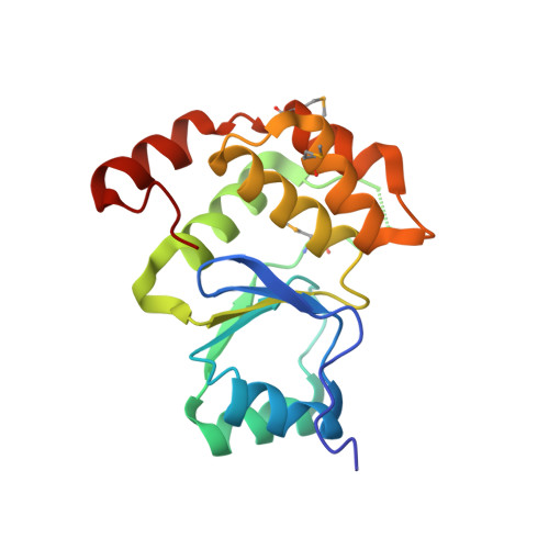
    Inositol pyrophosphates (PP-InsPs) are "energetic" intracellular signals that are ubiquitous in animals, plants, and fungi; structural and biochemical characterization of PP-InsP metabolic enzymes provides insight into their evolution, reaction mechanisms, and regulation. Here, we describe the 2.35-Å-resolution structure of the catalytic core of Siw14, a 5-PP-InsP phosphatase from Saccharomyces cerevisiae and a member of the protein tyrosine-phosphatase (PTP) superfamily. Conclusions that we derive from structural data are supported by extensive site-directed mutagenesis and kinetic analyses, thereby attributing new functional significance to several key residues. We demonstrate the high activity and exquisite specificity of Siw14 for the 5-diphosphate group of PP-InsPs. The three structural elements that demarcate a 9.2-Å-deep substrate-binding pocket each have spatial equivalents in PTPs, but we identify how these are specialized for Siw14 to bind and hydrolyze the intensely negatively charged PP-InsPs. ( a ) The catalytic P-loop with the C X 5 R(S/T) PTP motif contains additional, positively charged residues. ( b ) A loop between the α5 and α6 helices, corresponding to the Q-loop in PTPs, contains a lysine and an arginine that extend into the catalytic pocket due to displacement of the α5 helix orientation through intramolecular crowding caused by three bulky, hydrophobic residues. ( c ) The general-acid loop in PTPs is replaced in Siw14 with a flexible loop that does not use an aspartate or glutamate as a general acid. We propose that an acidic residue is not required for phosphoanhydride hydrolysis.


  • Organizational Affiliation

    From the Inositol Signaling Group, Signal Transduction Laboratory, NIEHS, National Institutes of Health, Research Triangle Park, North Carolina 27709, huanchen.wang@nih.gov.


Macromolecules
Find similar proteins by:  (by identity cutoff)  |  3D Structure
Entity ID: 1
MoleculeChains Sequence LengthOrganismDetailsImage
Tyrosine-protein phosphatase SIW14
A, B, C, D, E
A, B, C, D, E, F, G, H, I
170Saccharomyces cerevisiae S288CMutation(s): 0 
Gene Names: SIW14YNL032WN2746
EC: 3.1.3.48
UniProt
Find proteins for P53965 (Saccharomyces cerevisiae (strain ATCC 204508 / S288c))
Explore P53965 
Go to UniProtKB:  P53965
Entity Groups  
Sequence Clusters30% Identity50% Identity70% Identity90% Identity95% Identity100% Identity
UniProt GroupP53965
Sequence Annotations
Expand
  • Reference Sequence
Small Molecules
Ligands 3 Unique
IDChains Name / Formula / InChI Key2D Diagram3D Interactions
CIT
Query on CIT

Download Ideal Coordinates CCD File 
J [auth A]
L [auth B]
M [auth C]
O [auth D]
R [auth E]
J [auth A],
L [auth B],
M [auth C],
O [auth D],
R [auth E],
S [auth F],
T [auth G],
V [auth H],
W [auth I]
CITRIC ACID
C6 H8 O7
KRKNYBCHXYNGOX-UHFFFAOYSA-N
EDO
Query on EDO

Download Ideal Coordinates CCD File 
K [auth A],
N [auth C],
P [auth D]
1,2-ETHANEDIOL
C2 H6 O2
LYCAIKOWRPUZTN-UHFFFAOYSA-N
CL
Query on CL

Download Ideal Coordinates CCD File 
Q [auth D],
U [auth G]
CHLORIDE ION
Cl
VEXZGXHMUGYJMC-UHFFFAOYSA-M
Modified Residues  1 Unique
IDChains TypeFormula2D DiagramParent
MSE
Query on MSE
A, B, C, D, E
A, B, C, D, E, F, G, H, I
L-PEPTIDE LINKINGC5 H11 N O2 SeMET
Experimental Data & Validation

Experimental Data

  • Method: X-RAY DIFFRACTION
  • Resolution: 2.35 Å
  • R-Value Free: 0.229 
  • R-Value Work: 0.206 
  • R-Value Observed: 0.206 
  • Space Group: P 65 2 2
Unit Cell:
Length ( Å )Angle ( ˚ )
a = 92.968α = 90
b = 92.968β = 90
c = 814.716γ = 120
Software Package:
Software NamePurpose
PHENIXrefinement
HKL-2000data reduction
HKL-2000data scaling
AutoSolphasing

Structure Validation

View Full Validation Report


Entry History & Funding Information

Deposition Data


Funding OrganizationLocationGrant Number
National Institutes of Health/National Institute of Environmental Health Sciences (NIH/NIEHS)United States--

Revision History  (Full details and data files)

  • Version 1.0: 2018-03-21
    Type: Initial release
  • Version 1.1: 2018-03-28
    Changes: Data collection, Database references
  • Version 1.2: 2018-05-16
    Changes: Data collection, Database references
  • Version 1.3: 2022-03-16
    Changes: Author supporting evidence, Database references