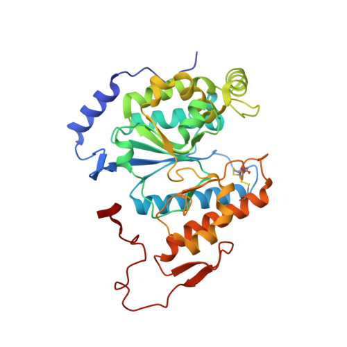
6BSV

Crystal structure of Xyloglucan Xylosyltransferase binary form


Experimental Data Snapshot

  • Method: X-RAY DIFFRACTION
  • Resolution: 2.43 Å
  • R-Value Free: 0.216 
  • R-Value Work: 0.184 
  • R-Value Observed: 0.186 

wwPDB Validation   3D Report Full Report


Ligand Structure Quality Assessment 


This is version 1.3 of the entry. See complete history


Literature

Structure of xyloglucan xylosyltransferase 1 reveals simple steric rules that define biological patterns of xyloglucan polymers.

Culbertson, A.T.Ehrlich, J.J.Choe, J.Y.Honzatko, R.B.Zabotina, O.A.

(2018) Proc Natl Acad Sci U S A 115: 6064-6069

  • DOI: https://doi.org/10.1073/pnas.1801105115
  • Primary Citation of Related Structures:  
    6BSU, 6BSV, 6BSW

  • PubMed Abstract: 

    The plant cell wall is primarily a polysaccharide mesh of the most abundant biopolymers on earth. Although one of the richest sources of biorenewable materials, the biosynthesis of the plant polysaccharides is poorly understood. Structures of many essential plant glycosyltransferases are unknown and suitable substrates are often unavailable for in vitro analysis. The dearth of such information impedes the development of plants better suited for industrial applications. Presented here are structures of Arabidopsis xyloglucan xylosyltransferase 1 (XXT1) without ligands and in complexes with UDP and cellohexaose. XXT1 initiates side-chain extensions from a linear glucan polymer by transferring the xylosyl group from UDP-xylose during xyloglucan biosynthesis. XXT1, a homodimer and member of the GT-A fold family of glycosyltransferases, binds UDP analogously to other GT-A fold enzymes. Structures here and the properties of mutant XXT1s are consistent with a SN i -like catalytic mechanism. Distinct from other systems is the recognition of cellohexaose by way of an extended cleft. The XXT1 dimer alone cannot produce xylosylation patterns observed for native xyloglucans because of steric constraints imposed by the acceptor binding cleft. Homology modeling of XXT2 and XXT5, the other two xylosyltransferases involved in xyloglucan biosynthesis, reveals a structurally altered cleft in XXT5 that could accommodate a partially xylosylated glucan chain produced by XXT1 and/or XXT2. An assembly of the three XXTs can produce the xylosylation patterns of native xyloglucans, suggesting the involvement of an organized multienzyme complex in the xyloglucan biosynthesis.


  • Organizational Affiliation

    Roy J. Carver Department of Biochemistry, Biophysics and Molecular Biology, Iowa State University, Ames, IA 50011.


Macromolecules
Find similar proteins by:  (by identity cutoff)  |  3D Structure
Entity ID: 1
MoleculeChains Sequence LengthOrganismDetailsImage
Xyloglucan 6-xylosyltransferase 1
A, B
338Arabidopsis thalianaMutation(s): 0 
Gene Names: XXT1XT1At3g62720F26K9_150
EC: 2.4.2.39
UniProt
Find proteins for Q9LZJ3 (Arabidopsis thaliana)
Explore Q9LZJ3 
Go to UniProtKB:  Q9LZJ3
Entity Groups  
Sequence Clusters30% Identity50% Identity70% Identity90% Identity95% Identity100% Identity
UniProt GroupQ9LZJ3
Sequence Annotations
Expand
  • Reference Sequence
Small Molecules
Ligands 4 Unique
IDChains Name / Formula / InChI Key2D Diagram3D Interactions
UDP
Query on UDP

Download Ideal Coordinates CCD File 
D [auth A],
H [auth B]
URIDINE-5'-DIPHOSPHATE
C9 H14 N2 O12 P2
XCCTYIAWTASOJW-XVFCMESISA-N
TRS
Query on TRS

Download Ideal Coordinates CCD File 
E [auth A],
I [auth B]
2-AMINO-2-HYDROXYMETHYL-PROPANE-1,3-DIOL
C4 H12 N O3
LENZDBCJOHFCAS-UHFFFAOYSA-O
NO3
Query on NO3

Download Ideal Coordinates CCD File 
F [auth A],
J [auth B]
NITRATE ION
N O3
NHNBFGGVMKEFGY-UHFFFAOYSA-N
MN
Query on MN

Download Ideal Coordinates CCD File 
C [auth A],
G [auth B]
MANGANESE (II) ION
Mn
WAEMQWOKJMHJLA-UHFFFAOYSA-N
Experimental Data & Validation

Experimental Data

  • Method: X-RAY DIFFRACTION
  • Resolution: 2.43 Å
  • R-Value Free: 0.216 
  • R-Value Work: 0.184 
  • R-Value Observed: 0.186 
  • Space Group: P 21 21 21
Unit Cell:
Length ( Å )Angle ( ˚ )
a = 64.929α = 90
b = 94.549β = 90
c = 144.716γ = 90
Software Package:
Software NamePurpose
PHENIXrefinement
PDB_EXTRACTdata extraction
HKL-3000data reduction
HKL-3000data scaling
PHENIXphasing

Structure Validation

View Full Validation Report


Ligand Structure Quality Assessment 


Entry History & Funding Information

Deposition Data


Funding OrganizationLocationGrant Number
National Science Foundation (NSF, United States)United States1121163

Revision History  (Full details and data files)

  • Version 1.0: 2018-05-23
    Type: Initial release
  • Version 1.1: 2018-06-06
    Changes: Data collection, Database references
  • Version 1.2: 2018-06-20
    Changes: Data collection, Database references
  • Version 1.3: 2022-03-23
    Changes: Advisory, Author supporting evidence, Database references, Derived calculations