6BPZ

Structure of the mechanically activated ion channel Piezo1

  • Classification: MEMBRANE PROTEIN
  • Organism(s): Mus musculus
  • Expression System: Homo sapiens
  • Mutation(s): No 
  • Membrane Protein: Yes  OPMPDBTM

  • Deposited: 2017-11-27 Released: 2017-12-27 
  • Deposition Author(s): Saotome, K., Kefauver, J.M., Patapoutian, A., Ward, A.B.
  • Funding Organization(s): Ray Thomas Edwards Foundation, National Institutes of Health/National Institute of Neurological Disorders and Stroke (NIH/NINDS), National Institutes of Health/National Institute of Dental and Craniofacial Research (NIH/NIDCR), Howard Hughes Medical Institute (HHMI)

Experimental Data Snapshot

  • Method: ELECTRON MICROSCOPY
  • Resolution: 3.80 Å
  • Aggregation State: PARTICLE 
  • Reconstruction Method: SINGLE PARTICLE 

wwPDB Validation   3D Report Full Report


This is version 1.5 of the entry. See complete history


Literature

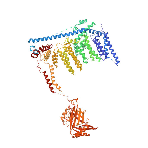
Structure of the mechanically activated ion channel Piezo1.

Saotome, K.Murthy, S.E.Kefauver, J.M.Whitwam, T.Patapoutian, A.Ward, A.B.

(2018) Nature 554: 481-486

  • DOI: https://doi.org/10.1038/nature25453
  • Primary Citation of Related Structures:  
    6BPZ

  • PubMed Abstract: 

    Piezo1 and Piezo2 are mechanically activated ion channels that mediate touch perception, proprioception and vascular development. Piezo proteins are distinct from other ion channels and their structure remains poorly defined, which impedes detailed study of their gating and ion permeation properties. Here we report a high-resolution cryo-electron microscopy structure of the mouse Piezo1 trimer. The detergent-solubilized complex adopts a three-bladed propeller shape with a curved transmembrane region containing at least 26 transmembrane helices per protomer. The flexible propeller blades can adopt distinct conformations, and consist of a series of four-transmembrane helical bundles that we term Piezo repeats. Carboxy-terminal domains line the central ion pore, and the channel is closed by constrictions in the cytosol. A kinked helical beam and anchor domain link the Piezo repeats to the pore, and are poised to control gating allosterically. The structure provides a foundation to dissect further how Piezo channels are regulated by mechanical force.


  • Organizational Affiliation

    Howard Hughes Medical Institute, Department of Neuroscience, The Scripps Research Institute, La Jolla, California 92037, USA.


Macromolecules
Find similar proteins by:  (by identity cutoff)  |  3D Structure
Entity ID: 1
MoleculeChains Sequence LengthOrganismDetailsImage
Piezo-type mechanosensitive ion channel component 1,Piezo-type mechanosensitive ion channel component 1,mouse Piezo1,Piezo-type mechanosensitive ion channel component 1,Piezo-type mechanosensitive ion channel component 1
A, B, C
1,423Mus musculusMutation(s): 0 
Gene Names: Piezo1Fam38a
Membrane Entity: Yes 
UniProt & NIH Common Fund Data Resources
Find proteins for E2JF22 (Mus musculus)
Explore E2JF22 
Go to UniProtKB:  E2JF22
IMPC:  MGI:3603204
Entity Groups  
Sequence Clusters30% Identity50% Identity70% Identity90% Identity95% Identity100% Identity
UniProt GroupE2JF22
Sequence Annotations
Expand
  • Reference Sequence
Experimental Data & Validation

Experimental Data

  • Method: ELECTRON MICROSCOPY
  • Resolution: 3.80 Å
  • Aggregation State: PARTICLE 
  • Reconstruction Method: SINGLE PARTICLE 
EM Software:
TaskSoftware PackageVersion
RECONSTRUCTIONRELION
MODEL REFINEMENTPHENIX

Structure Validation

View Full Validation Report


Entry History & Funding Information

Deposition Data


Funding OrganizationLocationGrant Number
Ray Thomas Edwards FoundationUnited States--
National Institutes of Health/National Institute of Neurological Disorders and Stroke (NIH/NINDS)United StatesNS083174
National Institutes of Health/National Institute of Dental and Craniofacial Research (NIH/NIDCR)United StatesDE022358
Howard Hughes Medical Institute (HHMI)United States--

Revision History  (Full details and data files)

  • Version 1.0: 2017-12-27
    Type: Initial release
  • Version 1.1: 2018-01-03
    Changes: Database references
  • Version 1.2: 2018-03-07
    Changes: Database references
  • Version 1.3: 2018-07-18
    Changes: Data collection, Experimental preparation
  • Version 1.4: 2019-11-20
    Changes: Author supporting evidence
  • Version 1.5: 2019-12-18
    Changes: Other