6BFG | pdb_00006bfg

Crystal structure of monotopic membrane protein (S)-mandelate dehydrogenase

  • Classification: OXIDOREDUCTASE
  • Organism(s): Pseudomonas putida
  • Expression System: Escherichia coli
  • Mutation(s): No 
  • Membrane Protein: Yes  OPMmpstruc

  • Deposited: 2017-10-26 Released: 2018-08-15 
  • Deposition Author(s): Sukumar, N.
  • Funding Organization(s): National Institutes of Health/National Institute of General Medical Sciences (NIH/NIGMS), National Institutes of Health/Office of the Director

Experimental Data Snapshot

  • Method: X-RAY DIFFRACTION
  • Resolution: 2.20 Å
  • R-Value Free: 
    0.240 (Depositor), 0.239 (DCC) 
  • R-Value Work: 
    0.197 (Depositor), 0.197 (DCC) 
  • R-Value Observed: 
    0.198 (Depositor) 

Starting Model: experimental
View more details

wwPDB Validation 3D Report Full Report

Validation slider image for 6BFG

Ligand Structure Quality Assessment 


This is version 1.4 of the entry. See complete history

Literature

Structure of the monotopic membrane protein (S)-mandelate dehydrogenase at 2.2 angstrom resolution.

Sukumar, N.Liu, S.Li, W.Mathews, F.S.Mitra, B.Kandavelu, P.

(2018) Biochimie 154: 45-54

  • DOI: https://doi.org/10.1016/j.biochi.2018.07.017
  • Primary Citation Related Structures: 
    6BFG

  • PubMed Abstract: 

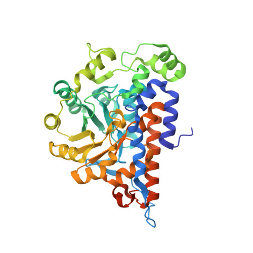
    The x-ray structure of the monotopic membrane protein (S)-mandelate dehydrogenase (MDH) from Pseudomonas putida reveals an inherent flexibility of its membrane binding segment that might be important for its biological activity. The surface of MDH exhibits a concentration of the positive charges on one side and the negative charges on the other side. The putative membrane binding surface of MDH has a concentric circular ridge, formed by positively charged residues, which projects away from the protein surface by ∼4 Å; this is an unique structural feature and not observed in other monotopic membrane proteins to our knowledge. There are three α-helixes in the membrane binding region. Based on the structure of MDH, it is possible to propose that the interaction of MDH with the membrane is stabilized by coplanar electrostatic interactions, between the positively charged concentric circular ridge and the negatively charged head-groups of the phospholipid bilayer, along with three α-helixes that provide additional stability by inserting into the membrane. The structure reveals the possible orientation of these helixes along with possible roles for the individual residues which form those helixes. These α-helixes may play a role in the enzyme's mobility. A detergent molecule, N-Dodecyl-β-maltoside, is inserted between the membrane binding region and rest of the molecule and may provide structural stability to intra-protein regions by forming hydrogen bonds and close contacts. From the average B-factor of the MDH structure, it is likely that MDH is highly mobile, which might be essential for its interaction in membrane and non-membrane environments, as its substrate (S)-mandelate, is from the cytoplasm, while its electron acceptor is a component of the membrane electron transport chain.


  • Organizational Affiliation
    • NE-CAT, Department of Chemistry and Chemical Biology, Cornell University, Building 436E, Argonne National Laboratory, Argonne, IL 60439, USA. Electronic address: sukumar@anl.gov.

Macromolecule Content 

  • Total Structure Weight: 91.38 kDa 
  • Atom Count: 6,264 
  • Modeled Residue Count: 746 
  • Deposited Residue Count: 798 
  • Unique protein chains: 1

Macromolecules

Find similar proteins by:|  3D Structure
Entity ID: 1
MoleculeChains  Sequence LengthOrganismDetailsImage
(S)-mandelate dehydrogenase
A, B
399Pseudomonas putidaMutation(s): 0 
Gene Names: mdlB
EC: 1.1.99.31
Membrane Entity: Yes 
UniProt
Find proteins for P20932 (Pseudomonas putida)
Explore P20932 
Go to UniProtKB:  P20932
Entity Groups
Sequence Clusters30% Identity50% Identity70% Identity90% Identity95% Identity100% Identity
UniProt GroupP20932
Sequence Annotations
Expand
Reference Sequence

Small Molecules

Ligands 9 Unique
IDChains Name / Formula / InChI Key2D Diagram3D Interactions
LMT

Query on LMT


Download:Ideal Coordinates CCD File
T [auth B]DODECYL-BETA-D-MALTOSIDE
C24 H46 O11
NLEBIOOXCVAHBD-QKMCSOCLSA-N
FMN

Query on FMN


Download:Ideal Coordinates CCD File
C [auth A],
N [auth B]
FLAVIN MONONUCLEOTIDE
C17 H21 N4 O9 P
FVTCRASFADXXNN-SCRDCRAPSA-N
CIT

Query on CIT


Download:Ideal Coordinates CCD File
L [auth A]CITRIC ACID
C6 H8 O7
KRKNYBCHXYNGOX-UHFFFAOYSA-N
PEG

Query on PEG


Download:Ideal Coordinates CCD File
O [auth B]DI(HYDROXYETHYL)ETHER
C4 H10 O3
MTHSVFCYNBDYFN-UHFFFAOYSA-N
SO4

Query on SO4


Download:Ideal Coordinates CCD File
G [auth A]SULFATE ION
O4 S
QAOWNCQODCNURD-UHFFFAOYSA-L
PO4

Query on PO4


Download:Ideal Coordinates CCD File
Q [auth B]PHOSPHATE ION
O4 P
NBIIXXVUZAFLBC-UHFFFAOYSA-K
GOL

Query on GOL


Download:Ideal Coordinates CCD File
D [auth A],
E [auth A],
F [auth A],
P [auth B]
GLYCEROL
C3 H8 O3
PEDCQBHIVMGVHV-UHFFFAOYSA-N
EDO

Query on EDO


Download:Ideal Coordinates CCD File
H [auth A]
I [auth A]
J [auth A]
K [auth A]
M [auth A]
H [auth A],
I [auth A],
J [auth A],
K [auth A],
M [auth A],
S [auth B],
U [auth B]
1,2-ETHANEDIOL
C2 H6 O2
LYCAIKOWRPUZTN-UHFFFAOYSA-N
NA

Query on NA


Download:Ideal Coordinates CCD File
R [auth B]SODIUM ION
Na
FKNQFGJONOIPTF-UHFFFAOYSA-N

Experimental Data & Validation

Experimental Data

  • Method: X-RAY DIFFRACTION
  • Resolution: 2.20 Å
  • R-Value Free:  0.240 (Depositor), 0.239 (DCC) 
  • R-Value Work:  0.197 (Depositor), 0.197 (DCC) 
  • R-Value Observed: 0.198 (Depositor) 
Space Group: I 2 2 2
Unit Cell:
Length ( Å )Angle ( ˚ )
a = 123.358α = 90
b = 129.879β = 90
c = 143.26γ = 90
Software Package:
Software NamePurpose
PHENIXrefinement
PDB_EXTRACTdata extraction
HKL-2000data reduction
HKL-2000data scaling
PHENIXphasing

Structure Validation

View Full Validation Report


Ligand Structure Quality Assessment 


Entry History 

& Funding Information

Deposition Data

  • Released Date: 2018-08-15 
  • Deposition Author(s): Sukumar, N.

Funding OrganizationLocationGrant Number
National Institutes of Health/National Institute of General Medical Sciences (NIH/NIGMS)United StatesGM103403
National Institutes of Health/Office of the DirectorUnited StatesS10OD021527

Revision History  (Full details and data files)

  • Version 1.0: 2018-08-15
    Type: Initial release
  • Version 1.1: 2018-11-07
    Changes: Data collection, Database references
  • Version 1.2: 2019-02-20
    Changes: Author supporting evidence, Data collection
  • Version 1.3: 2020-01-01
    Changes: Author supporting evidence
  • Version 1.4: 2023-10-04
    Changes: Advisory, Data collection, Database references, Refinement description