6BC8

Crystal structure of Rev7-R124A/Rev3-RBM2 (residues 1988-2014) complex

  • Classification: REPLICATION
  • Organism(s): Homo sapiens
  • Expression System: Escherichia coli BL21(DE3)
  • Mutation(s): Yes 

  • Deposited: 2017-10-20 Released: 2018-08-01 
  • Deposition Author(s): Rizzo, A.A., Hao, B., Li, Y., Korzhnev, D.M.
  • Funding Organization(s): National Science Foundation (NSF, United States), National Institutes of Health/National Institute of General Medical Sciences (NIH/NIGMS), National Institutes of Health/National Institute of Environmental Health Sciences (NIH/NIEHS), Connecticut Regenerative Medicine Research Fund

Experimental Data Snapshot

  • Method: X-RAY DIFFRACTION
  • Resolution: 1.68 Å
  • R-Value Free: 0.223 
  • R-Value Work: 0.181 
  • R-Value Observed: 0.183 

wwPDB Validation   3D Report Full Report


This is version 1.6 of the entry. See complete history


Literature

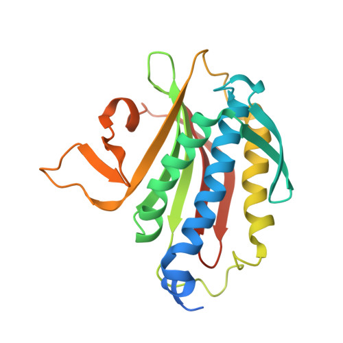
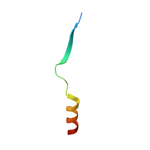
Rev7 dimerization is important for assembly and function of the Rev1/Pol zeta translesion synthesis complex.

Rizzo, A.A.Vassel, F.M.Chatterjee, N.D'Souza, S.Li, Y.Hao, B.Hemann, M.T.Walker, G.C.Korzhnev, D.M.

(2018) Proc Natl Acad Sci U S A 115: E8191-E8200

  • DOI: https://doi.org/10.1073/pnas.1801149115
  • Primary Citation of Related Structures:  
    6BC8, 6BCD, 6BI7

  • PubMed Abstract: 

    The translesion synthesis (TLS) polymerases Polζ and Rev1 form a complex that enables replication of damaged DNA. The Rev7 subunit of Polζ, which is a multifaceted HORMA (Hop1, Rev7, Mad2) protein with roles in TLS, DNA repair, and cell-cycle control, facilitates assembly of this complex by binding Rev1 and the catalytic subunit of Polζ, Rev3. Rev7 interacts with Rev3 by a mechanism conserved among HORMA proteins, whereby an open-to-closed transition locks the ligand underneath the "safety belt" loop. Dimerization of HORMA proteins promotes binding and release of this ligand, as exemplified by the Rev7 homolog, Mad2. Here, we investigate the dimerization of Rev7 when bound to the two Rev7-binding motifs (RBMs) in Rev3 by combining in vitro analyses of Rev7 structure and interactions with a functional assay in a Rev7 -/- cell line. We demonstrate that Rev7 uses the conventional HORMA dimerization interface both to form a homodimer when tethered by the two RBMs in Rev3 and to heterodimerize with other HORMA domains, Mad2 and p31 comet Structurally, the Rev7 dimer can bind only one copy of Rev1, revealing an unexpected Rev1/Polζ architecture. In cells, mutation of the Rev7 dimer interface increases sensitivity to DNA damage. These results provide insights into the structure of the Rev1/Polζ TLS assembly and highlight the function of Rev7 homo- and heterodimerization.


  • Organizational Affiliation

    Department of Molecular Biology and Biophysics, University of Connecticut Health Center, Farmington, CT 06030.


Macromolecules
Find similar proteins by:  (by identity cutoff)  |  3D Structure
Entity ID: 1
MoleculeChains Sequence LengthOrganismDetailsImage
Mitotic spindle assembly checkpoint protein MAD2B227Homo sapiensMutation(s): 1 
Gene Names: MAD2L2MAD2BREV7
UniProt & NIH Common Fund Data Resources
Find proteins for Q9UI95 (Homo sapiens)
Explore Q9UI95 
Go to UniProtKB:  Q9UI95
PHAROS:  Q9UI95
GTEx:  ENSG00000116670 
Entity Groups  
Sequence Clusters30% Identity50% Identity70% Identity90% Identity95% Identity100% Identity
UniProt GroupQ9UI95
Sequence Annotations
Expand
  • Reference Sequence
Find similar proteins by:  (by identity cutoff)  |  3D Structure
Entity ID: 2
MoleculeChains Sequence LengthOrganismDetailsImage
DNA polymerase zeta catalytic subunit28Homo sapiensMutation(s): 0 
Gene Names: REV3LPOLZREV3
EC: 2.7.7.7
UniProt & NIH Common Fund Data Resources
Find proteins for O60673 (Homo sapiens)
Explore O60673 
Go to UniProtKB:  O60673
PHAROS:  O60673
GTEx:  ENSG00000009413 
Entity Groups  
Sequence Clusters30% Identity50% Identity70% Identity90% Identity95% Identity100% Identity
UniProt GroupO60673
Sequence Annotations
Expand
  • Reference Sequence
Experimental Data & Validation

Experimental Data

  • Method: X-RAY DIFFRACTION
  • Resolution: 1.68 Å
  • R-Value Free: 0.223 
  • R-Value Work: 0.181 
  • R-Value Observed: 0.183 
  • Space Group: P 31 2 1
Unit Cell:
Length ( Å )Angle ( ˚ )
a = 63.691α = 90
b = 63.691β = 90
c = 116.274γ = 120
Software Package:
Software NamePurpose
REFMACrefinement
PHASERphasing
Cootmodel building
SCALEPACKdata scaling
DENZOdata reduction

Structure Validation

View Full Validation Report


Entry History & Funding Information

Deposition Data


Funding OrganizationLocationGrant Number
National Science Foundation (NSF, United States)United States1615866
National Institutes of Health/National Institute of General Medical Sciences (NIH/NIGMS)United StatesR01-GM123239
National Institutes of Health/National Institute of Environmental Health Sciences (NIH/NIEHS)United StatesR01-ES015818
National Institutes of Health/National Institute of Environmental Health Sciences (NIH/NIEHS)United StatesP30-ES002109
National Institutes of Health/National Institute of General Medical Sciences (NIH/NIGMS)United StatesGM099948
Connecticut Regenerative Medicine Research FundUnited States15-RMA- UCHC-03
National Science Foundation (NSF, United States)United StatesDMR-1332208

Revision History  (Full details and data files)

  • Version 1.0: 2018-08-01
    Type: Initial release
  • Version 1.1: 2018-08-22
    Changes: Data collection, Database references, Source and taxonomy
  • Version 1.2: 2018-08-29
    Changes: Data collection, Database references
  • Version 1.3: 2018-09-12
    Changes: Data collection, Database references
  • Version 1.4: 2018-09-19
    Changes: Data collection, Database references
  • Version 1.5: 2019-11-27
    Changes: Author supporting evidence
  • Version 1.6: 2023-10-04
    Changes: Data collection, Database references, Refinement description