Tilted, Uninterrupted, Monomeric HIV-1 gp41 Transmembrane Helix from Residual Dipolar Couplings.
Chiliveri, S.C., Louis, J.M., Ghirlando, R., Baber, J.L., Bax, A.(2018) J Am Chem Soc 140: 34-37
- PubMed: 29277995 Search on PubMedSearch on PubMed Central
- DOI: https://doi.org/10.1021/jacs.7b10245
- Primary Citation Related Structures:
6B3U - PubMed Abstract:
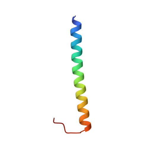
Cryo-electron microscopy and X-ray crystallography have shown that the pre- and postfusion states of the HIV-1 gp41 viral coat protein, although very different from one another, each adopt C 3 symmetric structures. A stable homotrimeric structure for the transmembrane domain (TM) also was modeled and supported by experimental data. For a C 3 symmetric structure, alignment in an anisotropic medium must be axially symmetric, with the unique axis of the alignment tensor coinciding with the C 3 axis. However, NMR residual dipolar couplings (RDCs) measured under three different alignment conditions were found to be incompatible with C 3 symmetry. Subsequent measurements by paramagnetic relaxation enhancement, analytical ultracentrifugation, and DEER EPR, indicate that the transmembrane domain is monomeric. 15 N NMR relaxation data and RDCs show that TM is highly ordered and uninterrupted for a total length of 32 residues, extending well into the membrane proximal external region.
- Laboratory of Chemical Physics and ‡Laboratory of Molecular Biology, National Institute of Diabetes and Digestive and Kidney Diseases, National Institutes of Health , Bethesda, Maryland 20892, United States.
Organizational Affiliation: