6AZ2 | pdb_00006az2

Crystal structure of Asf1-Fab 12E complex


Experimental Data Snapshot

  • Method: X-RAY DIFFRACTION
  • Resolution: 2.48 Å
  • R-Value Free: 
    0.228 (Depositor), 0.229 (DCC) 
  • R-Value Work: 
    0.193 (Depositor), 0.195 (DCC) 
  • R-Value Observed: 
    0.195 (Depositor) 

wwPDB Validation 3D Report Full Report

Validation slider image for 6AZ2

This is version 1.2 of the entry. See complete history

Literature

Locking the Elbow: Improved Antibody Fab Fragments as Chaperones for Structure Determination.

Bailey, L.J.Sheehy, K.M.Dominik, P.K.Liang, W.G.Rui, H.Clark, M.Jaskolowski, M.Kim, Y.Deneka, D.Tang, W.J.Kossiakoff, A.A.

(2018) J Mol Biology 430: 337-347

  • DOI: https://doi.org/10.1016/j.jmb.2017.12.012
  • Primary Citation Related Structures: 
    5CJO, 6AYZ, 6AZ2

  • PubMed Abstract: 

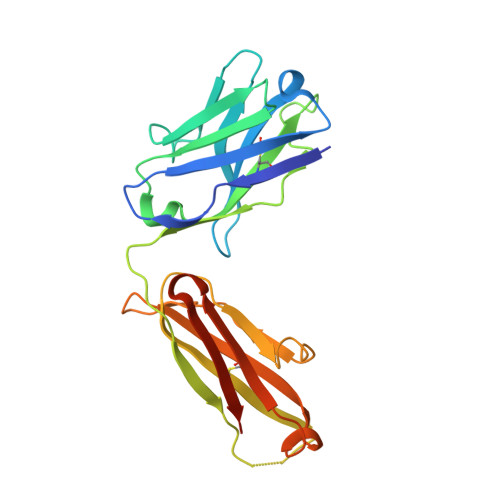
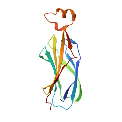
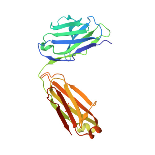
    Antibody Fab fragments have been exploited with significant success to facilitate the structure determination of challenging macromolecules as crystallization chaperones and as molecular fiducial marks for single particle cryo-electron microscopy approaches. However, the inherent flexibility of the "elbow" regions, which link the constant and variable domains of the Fab, can introduce disorder and thus diminish their effectiveness. We have developed a phage display engineering strategy to generate synthetic Fab variants that significantly reduces elbow flexibility, while maintaining their high affinity and stability. This strategy was validated using previously recalcitrant Fab-antigen complexes where introduction of an engineered elbow region enhanced crystallization and diffraction resolution. Furthermore, incorporation of the mutations appears to be generally portable to other synthetic antibodies and may serve as a universal strategy to enhance the success rates of Fabs as structure determination chaperones.


  • Organizational Affiliation
    • Department of Biochemistry and Molecular Biology, University of Chicago, Chicago, IL 60637, USA.

Macromolecule Content 

  • Total Structure Weight: 130.59 kDa 
  • Atom Count: 8,950 
  • Modeled Residue Count: 1,131 
  • Deposited Residue Count: 1,196 
  • Unique protein chains: 3

Macromolecules

Find similar proteins by:|  3D Structure
Entity ID: 1
MoleculeChains  Sequence LengthOrganismDetailsImage
Fab Heavy ChainA,
B [auth C]
229Homo sapiensMutation(s): 0 
Find similar proteins by:|  3D Structure
Entity ID: 2
MoleculeChains  Sequence LengthOrganismDetailsImage
Histone chaperone ASF1C [auth B],
D
154Saccharomyces cerevisiae S288CMutation(s): 0 
Gene Names: ASF1CIA1YJL115WJ0755
UniProt
Find proteins for P32447 (Saccharomyces cerevisiae (strain ATCC 204508 / S288c))
Explore P32447 
Go to UniProtKB:  P32447
Entity Groups
Sequence Clusters30% Identity50% Identity70% Identity90% Identity95% Identity100% Identity
UniProt GroupP32447
Sequence Annotations
Expand
Reference Sequence
Find similar proteins by:|  3D Structure
Entity ID: 3
MoleculeChains  Sequence LengthOrganismDetailsImage
Fab Light Chain
E, F
215Homo sapiensMutation(s): 0 

Experimental Data & Validation

Experimental Data

  • Method: X-RAY DIFFRACTION
  • Resolution: 2.48 Å
  • R-Value Free:  0.228 (Depositor), 0.229 (DCC) 
  • R-Value Work:  0.193 (Depositor), 0.195 (DCC) 
  • R-Value Observed: 0.195 (Depositor) 
Space Group: P 21 21 21
Unit Cell:
Length ( Å )Angle ( ˚ )
a = 99.666α = 90
b = 109.743β = 90
c = 122.17γ = 90
Software Package:
Software NamePurpose
PHENIXrefinement
HKL-3000data collection
SCALEPACKdata scaling
MOLREPphasing

Structure Validation

View Full Validation Report


Entry History 

Deposition Data

Revision History  (Full details and data files)

  • Version 1.0: 2018-01-10
    Type: Initial release
  • Version 1.1: 2018-02-14
    Changes: Database references
  • Version 1.2: 2024-11-13
    Changes: Data collection, Database references, Structure summary