6ASZ

Chromodomain HP1 with Y24F mutation bound to histone H3 peptide containing trimethyl lysine


Experimental Data Snapshot

  • Method: X-RAY DIFFRACTION
  • Resolution: 1.52 Å
  • R-Value Free: 0.274 
  • R-Value Work: 0.255 
  • R-Value Observed: 0.256 

wwPDB Validation   3D Report Full Report


This is version 1.3 of the entry. See complete history


Literature

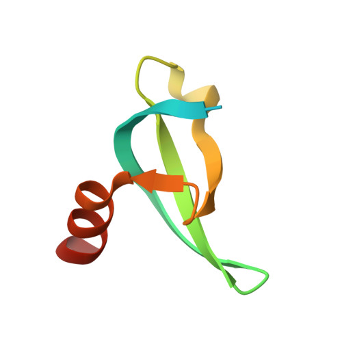
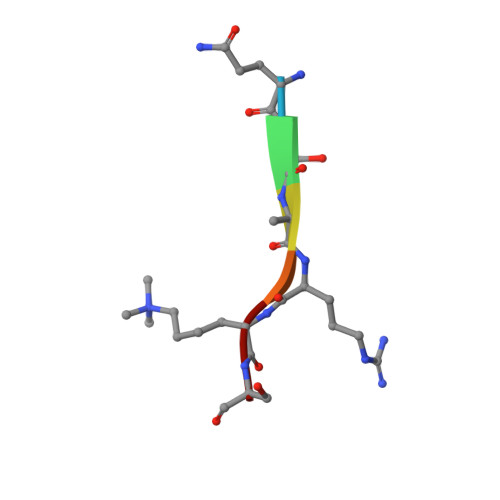
Investigation of Trimethyllysine Binding by the HP1 Chromodomain via Unnatural Amino Acid Mutagenesis.

Baril, S.A.Koenig, A.L.Krone, M.W.Albanese, K.I.He, C.Q.Lee, G.Y.Houk, K.N.Waters, M.L.Brustad, E.M.

(2017) J Am Chem Soc 139: 17253-17256

  • DOI: https://doi.org/10.1021/jacs.7b09223
  • Primary Citation of Related Structures:  
    6ASZ, 6AT0

  • PubMed Abstract: 

    Trimethyllysine (Kme3) reader proteins are targets for inhibition due to their role in mediating gene expression. Although all such reader proteins bind Kme3 in an aromatic cage, the driving force for binding may differ; some readers exhibit evidence for cation-π interactions whereas others do not. We report a general unnatural amino acid mutagenesis approach to quantify the contribution of individual tyrosines to cation binding using the HP1 chromodomain as a model system. We demonstrate that two tyrosines (Y24 and Y48) bind to a Kme3-histone tail peptide via cation-π interactions, but linear free energy trends suggest they do not contribute equally to binding. X-ray structures and computational analysis suggest that the distance and degree of contact between Tyr residues and Kme3 plays an important role in tuning cation-π-mediated Kme3 recognition. Although cation-π interactions have been studied in a number of proteins, this work is the first to utilize direct binding assays, X-ray crystallography, and modeling, to pinpoint factors that influence the magnitude of the individual cation-π interactions.


  • Organizational Affiliation

    Department of Chemistry, CB 3290, University of North Carolina at Chapel Hill , Chapel Hill, North Carolina 27599, United States.


Macromolecules
Find similar proteins by:  (by identity cutoff)  |  3D Structure
Entity ID: 1
MoleculeChains Sequence LengthOrganismDetailsImage
Heterochromatin protein 169Drosophila melanogasterMutation(s): 2 
Gene Names: Su(var)205HP1CG8409
UniProt
Find proteins for P05205 (Drosophila melanogaster)
Explore P05205 
Go to UniProtKB:  P05205
Entity Groups  
Sequence Clusters30% Identity50% Identity70% Identity90% Identity95% Identity100% Identity
UniProt GroupP05205
Sequence Annotations
Expand
  • Reference Sequence

Find similar proteins by:  Sequence   |   3D Structure  

Entity ID: 2
MoleculeChains Sequence LengthOrganismDetailsImage
trimethyl lysine histone H3 tail peptideB [auth P]6Drosophila melanogasterMutation(s): 0 
UniProt
Find proteins for P02299 (Drosophila melanogaster)
Explore P02299 
Go to UniProtKB:  P02299
Entity Groups  
UniProt GroupP02299
Sequence Annotations
Expand
  • Reference Sequence
Small Molecules
Modified Residues  1 Unique
IDChains TypeFormula2D DiagramParent
M3L
Query on M3L
B [auth P]L-PEPTIDE LINKINGC9 H21 N2 O2LYS
Experimental Data & Validation

Experimental Data

  • Method: X-RAY DIFFRACTION
  • Resolution: 1.52 Å
  • R-Value Free: 0.274 
  • R-Value Work: 0.255 
  • R-Value Observed: 0.256 
  • Space Group: C 2 2 21
Unit Cell:
Length ( Å )Angle ( ˚ )
a = 34.516α = 90
b = 76.78β = 90
c = 75.508γ = 90
Software Package:
Software NamePurpose
PHASERphasing
PHENIXrefinement
PDB_EXTRACTdata extraction

Structure Validation

View Full Validation Report


Entry History 

Deposition Data

Revision History  (Full details and data files)

  • Version 1.0: 2017-12-06
    Type: Initial release
  • Version 1.1: 2017-12-13
    Changes: Database references
  • Version 1.2: 2019-03-27
    Changes: Advisory, Data collection
  • Version 1.3: 2023-10-04
    Changes: Data collection, Database references, Refinement description