6AHH

Crystal Structure of HEWL in complex with Phenylethyl alcohol (in the aroma form) after 5 hours under fibrillation conditions


Experimental Data Snapshot

  • Method: X-RAY DIFFRACTION
  • Resolution: 2.10 Å
  • R-Value Free: 0.246 
  • R-Value Work: 0.178 
  • R-Value Observed: 0.181 

wwPDB Validation   3D Report Full Report


This is version 1.2 of the entry. See complete history


Literature

Cinnamaldehyde and Phenyl Ethyl Alcohol promote the entrapment of intermediate species of HEWL, as revealed by structural, kinetics and thermal stability studies.

Seraj, Z.Groves, M.R.Seyedarabi, A.

(2019) Sci Rep 9: 18615-18615

  • DOI: https://doi.org/10.1038/s41598-019-55082-1
  • Primary Citation of Related Structures:  
    6AC2, 6AHH, 6AHL

  • PubMed Abstract: 

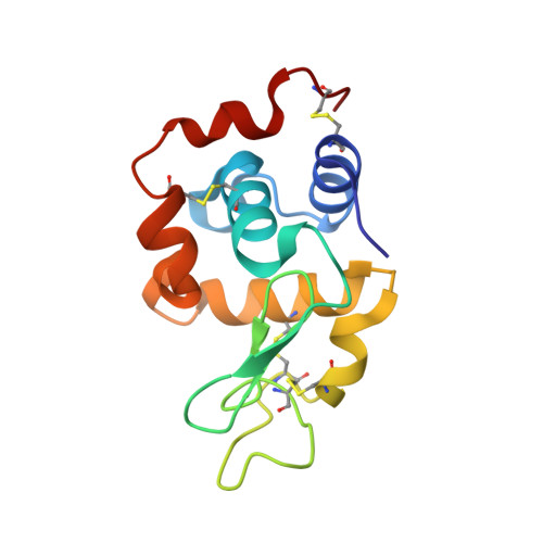
    Numerous efforts have been directed towards investigating the different stages leading to the fibrillation process in neurodegenerative diseases and finding the factors modulating it. In this study, using a wide range of molecular techniques as well as fibrillation kinetics coupled with differential scanning fluorimetry (DSF) and crystal structure determination of HEWL treated with cinnamaldehyde (Cin) and Phenyl ethyl alcohol (PEA) in their aroma form during fibrillation, we were able to identify the binding positions of Cin and PEA in HEWL. Additionally, crystal structures were used to suggest residues Thr43, Asn44, Arg45 and Arg68 as a plausible 'hotspot' promoting entrapment of intermediate species in the process of fibril formation in HEWL. We were also able to use DSF to show that Cin can significantly decrease the thermal stability of HEWL, promoting the formation of partially unfolded intermediate species. In conclusion, our data led us to emphasize that compounds in their 'aroma form' can influence the structure and stability of protein molecules and suggest reconsideration of HEWL as a model protein for fibrillation studies related to neurodegenerative diseases based on the initial structure of the proteins, whether globular (HEWL) or intrinsically disordered.


  • Organizational Affiliation

    Department of Biochemistry, Institute of Biochemistry and Biophysics, University of Tehran, Tehran, Iran.


Macromolecules
Find similar proteins by:  (by identity cutoff)  |  3D Structure
Entity ID: 1
MoleculeChains Sequence LengthOrganismDetailsImage
Lysozyme C129Gallus gallusMutation(s): 0 
EC: 3.2.1.17
UniProt
Find proteins for P00698 (Gallus gallus)
Explore P00698 
Go to UniProtKB:  P00698
Entity Groups  
Sequence Clusters30% Identity50% Identity70% Identity90% Identity95% Identity100% Identity
UniProt GroupP00698
Sequence Annotations
Expand
  • Reference Sequence
Small Molecules
Ligands 4 Unique
IDChains Name / Formula / InChI Key2D Diagram3D Interactions
PEL
Query on PEL

Download Ideal Coordinates CCD File 
F [auth A],
G [auth A]
2-PHENYL-ETHANOL
C8 H10 O
WRMNZCZEMHIOCP-UHFFFAOYSA-N
PGO
Query on PGO

Download Ideal Coordinates CCD File 
B [auth A]S-1,2-PROPANEDIOL
C3 H8 O2
DNIAPMSPPWPWGF-VKHMYHEASA-N
CL
Query on CL

Download Ideal Coordinates CCD File 
C [auth A],
D [auth A],
E [auth A]
CHLORIDE ION
Cl
VEXZGXHMUGYJMC-UHFFFAOYSA-M
NA
Query on NA

Download Ideal Coordinates CCD File 
H [auth A]SODIUM ION
Na
FKNQFGJONOIPTF-UHFFFAOYSA-N
Experimental Data & Validation

Experimental Data

  • Method: X-RAY DIFFRACTION
  • Resolution: 2.10 Å
  • R-Value Free: 0.246 
  • R-Value Work: 0.178 
  • R-Value Observed: 0.181 
  • Space Group: P 43 21 2
Unit Cell:
Length ( Å )Angle ( ˚ )
a = 78.667α = 90
b = 78.667β = 90
c = 37.382γ = 90
Software Package:
Software NamePurpose
REFMACrefinement
XDSdata reduction
autoPROCdata scaling
SCALAdata scaling
PHASERphasing

Structure Validation

View Full Validation Report


Entry History 

Deposition Data

Revision History  (Full details and data files)

  • Version 1.0: 2019-08-28
    Type: Initial release
  • Version 1.1: 2020-01-29
    Changes: Database references
  • Version 1.2: 2023-11-22
    Changes: Data collection, Database references, Refinement description