6AEI | pdb_00006aei

Cryo-EM structure of the receptor-activated TRPC5 ion channel


Experimental Data Snapshot

  • Method: ELECTRON MICROSCOPY
  • Resolution: 2.89 Å
  • Aggregation State: PARTICLE 
  • Reconstruction Method: SINGLE PARTICLE 

wwPDB Validation 3D Report Full Report

Validation slider image for 6AEI

This is version 1.3 of the entry. See complete history

Literature

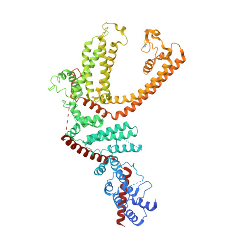
Cryo-EM structure of TRPC5 at 2.8- angstrom resolution reveals unique and conserved structural elements essential for channel function.

Duan, J.Li, J.Chen, G.L.Ge, Y.Liu, J.Xie, K.Peng, X.Zhou, W.Zhong, J.Zhang, Y.Xu, J.Xue, C.Liang, B.Zhu, L.Liu, W.Zhang, C.Tian, X.L.Wang, J.Clapham, D.E.Zeng, B.Li, Z.Zhang, J.

(2019) Sci Adv 5

  • DOI: https://doi.org/10.1126/sciadv.aaw7935
  • Primary Citation Related Structures: 
    6AEI

  • PubMed Abstract: 

    The transient receptor potential canonical subfamily member 5 (TRPC5), one of seven mammalian TRPC members, is a nonselective calcium-permeant cation channel. TRPC5 is of considerable interest as a drug target in the treatment of progressive kidney disease, depression, and anxiety. Here, we present the 2.8-Å resolution cryo-electron microscopy (cryo-EM) structure of the mouse TRPC5 (mTRPC5) homotetramer. Comparison of the TRPC5 structure to previously determined structures of other TRPC and TRP channels reveals differences in the extracellular pore domain and in the length of the S3 helix. The disulfide bond at the extracellular side of the pore and a preceding small loop are essential elements for its proper function. This high-resolution structure of mTRPC5, combined with electrophysiology and mutagenesis, provides insight into the lipid modulation and gating mechanisms of the TRPC family of ion channels.


  • Organizational Affiliation
    • Human Aging Research Institute (HARI), School of Life Sciences, Nanchang University, Nanchang, Jiangxi 330031, China.

Macromolecule Content 

  • Total Structure Weight: 360.27 kDa 
  • Atom Count: 21,142 
  • Modeled Residue Count: 2,568 
  • Deposited Residue Count: 3,060 
  • Unique protein chains: 1

Macromolecules

Find similar proteins by:|  3D Structure
Entity ID: 1
MoleculeChains  Sequence LengthOrganismDetailsImage
Short transient receptor potential channel 5
A, B, C, D
765Mus musculusMutation(s): 0 
Gene Names: Trpc5Trp5Trrp5
Membrane Entity: Yes 
UniProt
Find proteins for Q9QX29 (Mus musculus)
Explore Q9QX29 
Go to UniProtKB:  Q9QX29
Entity Groups
Sequence Clusters30% Identity50% Identity70% Identity90% Identity95% Identity100% Identity
UniProt GroupQ9QX29
Sequence Annotations
Expand
Reference Sequence

Small Molecules

Ligands 3 Unique
IDChains Name / Formula / InChI Key2D Diagram3D Interactions
LPP

Query on LPP


Download:Ideal Coordinates CCD File
H [auth A],
K [auth B],
N [auth C],
Q [auth D]
2-(HEXADECANOYLOXY)-1-[(PHOSPHONOOXY)METHYL]ETHYL HEXADECANOATE
C35 H69 O8 P
PORPENFLTBBHSG-MGBGTMOVSA-N
Y01

Query on Y01


Download:Ideal Coordinates CCD File
G [auth A],
J [auth B],
M [auth C],
P [auth D]
CHOLESTEROL HEMISUCCINATE
C31 H50 O4
WLNARFZDISHUGS-MIXBDBMTSA-N
NA

Query on NA


Download:Ideal Coordinates CCD File
E [auth A],
F [auth A],
I [auth B],
L [auth C],
O [auth D]
SODIUM ION
Na
FKNQFGJONOIPTF-UHFFFAOYSA-N

Experimental Data & Validation

Experimental Data

  • Method: ELECTRON MICROSCOPY
  • Resolution: 2.89 Å
  • Aggregation State: PARTICLE 
  • Reconstruction Method: SINGLE PARTICLE 
EM Software:
TaskSoftware PackageVersion
MODEL REFINEMENTPHENIX

Structure Validation

View Full Validation Report


Entry History 

Deposition Data

Revision History  (Full details and data files)

  • Version 1.0: 2019-08-07
    Type: Initial release
  • Version 1.1: 2019-08-14
    Changes: Data collection, Database references
  • Version 1.2: 2024-10-16
    Changes: Data collection, Database references, Structure summary
  • Version 1.3: 2025-06-18
    Changes: Data collection