Multicolor redox sensor proteins can visualize redox changes in various compartments of the living cell.
Sugiura, K., Tanaka, H., Kurisu, G., Wakabayashi, K.I., Hisabori, T.(2019) Biochim Biophys Acta Gen Subj 1863: 1098-1107
- PubMed: 30953671 Search on PubMed
- DOI: https://doi.org/10.1016/j.bbagen.2019.01.016
- Primary Citation Related Structures:
6AA2, 6AA6 - PubMed Abstract:
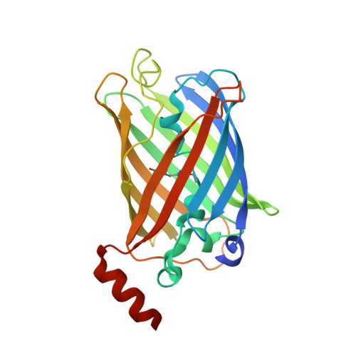
Change in the intracellular redox state is a consequence of various metabolic reactions, which simultaneously regulates various physiological phenomena in cells. Monitoring the redox state in living cells is thus very important for understanding cellular physiology. Various genetically encoded fluorescent redox sensors have therefore been developed. Recently, we developed oxidation-sensitive fluorescent proteins named Oba-Q (Sugiura, K., et al. (2015) Biochem. Biophys. Res. Commun. 457, 242-248), which exhibit dramatic quenching under oxidizing conditions. To extend the range of uses of redox sensor proteins, we refined these proteins based on the molecular architecture applied to Oba-Q, and successfully produced several redox sensor proteins based on CFP and YFP. Interestingly, some of these sensor proteins showed the reverse changes in emission compared with Oba-Q, implying remarkable fluorescence quenching under reducing conditions. We named this type of sensor protein Re-Q, reduction-sensed quenching protein. The cause of the redox-dependent fluorescence quenching could be clearly explained based on the crystal structure of Re-Q in the reduced and oxidized forms. In addition, by introducing suitable mutations into the sensors, we produced Oba-Q and Re-Q mutants exhibiting various midpoint redox potentials. This series of proteins can cover a wide range of redox potentials in the cell, so they should be applicable to various cells and even intracellular organelles. As an example, we successfully measured the redox responses in different cell compartments of cultured mammalian cells simultaneously against the anticancer reagents Kp372-1.
- Laboratory for Chemistry and Life Science, Institute of Innovative Research, Tokyo Institute of Technology, Nagatsuta-cho 4259, Midori-ku, Yokohama 226-8503, Japan; Core Research for Evolutional Science and Technology (CREST), Japan Science and Technology Agency (JST), Chiyoda-Ku, Tokyo 102-0076, Japan.
Organizational Affiliation: