Structural and mechanistic insights into polymyxin resistance mediated by EptC originating from Escherichia coli.
Zhao, Y., Meng, Q., Lai, Y., Wang, L., Zhou, D., Dou, C., Gu, Y., Nie, C., Wei, Y., Cheng, W.(2019) FEBS J 286: 750-764
- PubMed: 30537137 Search on PubMed
- DOI: https://doi.org/10.1111/febs.14719
- Primary Citation Related Structures:
6A82, 6A83 - PubMed Abstract:
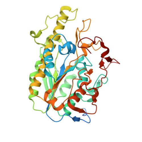
Gram-negative bacteria defend against the toxicity of polymyxins by modifying their outer membrane lipopolysaccharide (LPS). This modification mainly occurs through the addition of cationic molecules such as phosphoethanolamine (PEA). EcEptC is a PEA transferase from Escherichia coli (E. coli). However, unlike its homologs CjEptC (Campylobacter jejuni) and MCR-1, EcEptC is unable to mediate polymyxin resistance when overexpressed in E. coli. Here, we report crystal structures of the C-terminal putative catalytic domain (EcEptCΔN, 205-577 aa) of EcEptC in apo and Zn 2+ -bound states at 2.10 and 2.60 Å, respectively. EcEptCΔN is arranged into an α-β-α fold and equipped with the zinc ion in a conserved mode. Coupled with isothermal titration calorimetry (ITC) data, we provide insights into the mechanism by which EcEptC recognizes Zn 2+ . Furthermore, structure comparison analysis indicated that disulfide bonds, which play a key role in polymyxin resistance, were absent in EcEptCΔN. Supported by structural and biochemical evidence, we reveal mechanistic implications for disulfide bonds in PEA transferase-mediated polymyxin resistance. Significantly, because the structural effects exhibited by disulfide bonds are absent in EcEptC, it is impossible for this protein to participate in polymyxin resistance in E. coli. DATABASE: Structural data are available in the PDB under the accession numbers 6A82 and 6A83. ENZYME: EC 2.7.8.43.
- Division of Respiratory and Critical Care Medicine, State Key Laboratory of Biotherapy, West China Hospital of Sichuan University and Collaborative Innovation Center of Biotherapy, Chengdu, China.
Organizational Affiliation: