6A79 | pdb_00006a79

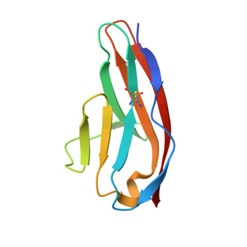
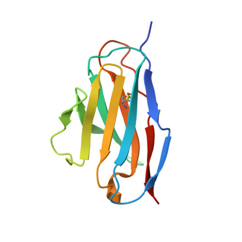
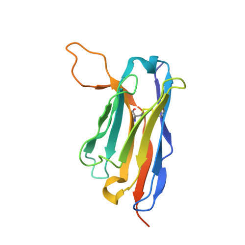
Crystal structure of the fifth immunoglobulin domain (Ig5) of human Robo1 in complex with the mutant scFv fragment (P103A) of murine monoclonal antibody B5209B


Experimental Data Snapshot

  • Method: X-RAY DIFFRACTION
  • Resolution: 2.31 Å
  • R-Value Free: 
    0.295 (Depositor), 0.242 (DCC) 
  • R-Value Work: 
    0.273 (Depositor) 
  • R-Value Observed: 
    0.274 (Depositor) 

wwPDB Validation 3D Report Full Report

Validation slider image for 6A79

This is version 1.2 of the entry. See complete history. 

Literature

Affinity Improvement of a Cancer-Targeted Antibody through Alanine-Induced Adjustment of Antigen-Antibody Interface.

Yamashita, T., Mizohata, E., Nagatoishi, S., Watanabe, T., Nakakido, M., Iwanari, H., Mochizuki, Y., Nakayama, T., Kado, Y., Yokota, Y., Matsumura, H., Kawamura, T., Kodama, T., Hamakubo, T., Inoue, T., Fujitani, H., Tsumoto, K.

(2019) Structure 27: 519

  • DOI: https://doi.org/10.1016/j.str.2018.11.002
  • Primary Citation Related Structures: 
    6A76, 6A77, 6A78, 6A79

  • PubMed Abstract: 

    To investigate favorable single amino acid substitutions that improve antigen-antibody interactions, alanine (Ala) mutagenesis scanning of the interfacial residues of a cancer-targeted antibody, B5209B, was performed based on X-ray crystallography analysis. Two substitutions were shown to significantly enhance the binding affinity for the antigen, by up to 30-fold. One substitution improved the affinity by a gain of binding enthalpy, whereas the other substitution improved the affinity by a gain of binding entropy. Molecular dynamics simulations showed that the enthalpic improvement could be attributed to the stabilization of distant salt bridges located at the periphery of the antigen-antibody interface. The entropic improvement was due to the release of water molecules that were stably trapped in the antigen-antibody interface of the wild-type antibody. Importantly, these effects of the Ala substitutions were caused by subtle adjustments of the binding interface. These results will be helpful to design high-affinity antibodies with avoiding entropy-enthalpy compensation.


  • Organizational Affiliation: 
    • Laboratory for Systems Biology and Medicine, Research Center for Advanced Science and Technology, The University of Tokyo, 4-6-1, Komaba, Meguro-ku, Tokyo 153-8904, Japan.

Macromolecule Content 

  • Total Structure Weight: 72.44 kDa 
  • Atom Count: 5,238 
  • Modeled Residue Count: 641 
  • Deposited Residue Count: 682 
  • Unique protein chains: 3

Macromolecules

Find similar proteins by:|  3D Structure
Entity ID: 1
MoleculeChains  Sequence LengthOrganismDetailsImage
Roundabout homolog 1A,
D [auth B]
91Homo sapiensMutation(s): 0 
Gene Names: ROBO1, DUTT1
UniProt & NIH Common Fund Data Resources
Find proteins for Q9Y6N7 (Homo sapiens)
Explore Q9Y6N7 
Go to UniProtKB:  Q9Y6N7
PHAROS:  Q9Y6N7
GTEx:  ENSG00000169855 
Entity Groups
Sequence Clusters30% Identity50% Identity70% Identity90% Identity95% Identity100% Identity
UniProt GroupQ9Y6N7
Sequence Annotations
Expand
Reference Sequence
Find similar proteins by:|  3D Structure
Entity ID: 2
MoleculeChains  Sequence LengthOrganismDetailsImage
Light chain region of the anti-human Robo1 antibody B5209B scFvB [auth L],
E [auth M]
112Mus musculusMutation(s): 0 
Entity Groups
Sequence Clusters30% Identity50% Identity70% Identity90% Identity95% Identity100% Identity
Sequence Annotations
Expand
Reference Sequence
Find similar proteins by:|  3D Structure
Entity ID: 3
MoleculeChains  Sequence LengthOrganismDetailsImage
Heavy chain of the anti-human Robo1 antibody B5209B scFvC [auth H],
F [auth I]
138Mus musculusMutation(s): 0 
Entity Groups
Sequence Clusters30% Identity50% Identity70% Identity90% Identity95% Identity100% Identity
Sequence Annotations
Expand
Reference Sequence

Experimental Data & Validation

Experimental Data

  • Method: X-RAY DIFFRACTION
  • Resolution: 2.31 Å
  • R-Value Free:  0.295 (Depositor), 0.242 (DCC) 
  • R-Value Work:  0.273 (Depositor) 
  • R-Value Observed: 0.274 (Depositor) 
Space Group: P 21 21 2
Unit Cell:
Length ( Å )Angle ( ˚ )
a = 70.766α = 90
b = 150.219β = 90
c = 66.852γ = 90
Software Package:
Software NamePurpose
REFMACrefinement
HKL-2000data reduction
Cootmodel building
PHASERphasing

Structure Validation

View Full Validation Report


Entry History 

& Funding Information

Deposition Data


Funding OrganizationLocationGrant Number
JapanFIRST Program (MDADD)

Revision History  (Full details and data files)

  • Version 1.0: 2019-01-30
    Type: Initial release
  • Version 1.1: 2019-03-20
    Changes: Data collection, Database references
  • Version 1.2: 2024-10-30
    Changes: Data collection, Database references, Structure summary