5ZNN

Crystal structure of ligand-free form of the Vps10 ectodomain of Sortilin


Experimental Data Snapshot

  • Method: X-RAY DIFFRACTION
  • Resolution: 2.45 Å
  • R-Value Free: 0.217 
  • R-Value Work: 0.180 
  • R-Value Observed: 0.181 

wwPDB Validation   3D Report Full Report


Ligand Structure Quality Assessment 


This is version 2.1 of the entry. See complete history


Literature

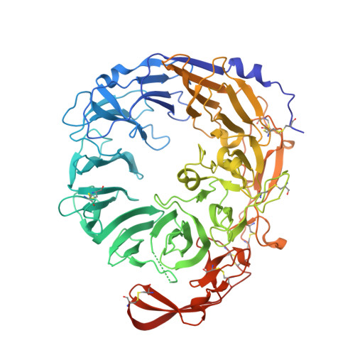
Crystal structure of the ligand-free form of the Vps10 ectodomain of dimerized Sortilin at acidic pH

Yabe-Wada, T.Matsuba, S.Unno, M.Onai, N.

(2018) FEBS Lett 592: 2647-2657

  • DOI: https://doi.org/10.1002/1873-3468.13181
  • Primary Citation of Related Structures:  
    5ZNN

  • PubMed Abstract: 

    Sortilin is a multifunctional sorting receptor involved in cytokine production in immune cells. To understand the mechanism of Sortilin-mediated cytokine trafficking, we determined the 2.45-Å structure of the dimerized Sortilin ectodomain (sSortilin or the Vps10-domain) crystallized at acidic pH. Substantial conformational changes upon dimerization lead to the intermolecular hydrophobic interaction between the conserved E455 and F137. Analysis of the electrostatic surface and size-exclusion chromatography revealed that sSortilin dimerization occurs due to an increase in hydrophobic interactions at the neutral dimer interface at acidic pH. The N682-attached N-glycan in the vicinity of the dimer interface implies its involvement in the dimerization. The disruption of Sortilin dimerization by mutations impairs efficient interferon-alpha secretion from cells. These results suggest the functional importance of Sortilin dimerization in cytokine trafficking.


  • Organizational Affiliation

    Department of Immunology, Kanazawa Medical University, Uchinada, Japan.


Macromolecules
Find similar proteins by:  (by identity cutoff)  |  3D Structure
Entity ID: 1
MoleculeChains Sequence LengthOrganismDetailsImage
Sortilin
A, B
679Mus musculusMutation(s): 0 
Gene Names: Sort1
UniProt & NIH Common Fund Data Resources
Find proteins for Q6PHU5 (Mus musculus)
Explore Q6PHU5 
Go to UniProtKB:  Q6PHU5
IMPC:  MGI:1338015
Entity Groups  
Sequence Clusters30% Identity50% Identity70% Identity90% Identity95% Identity100% Identity
UniProt GroupQ6PHU5
Sequence Annotations
Expand
  • Reference Sequence
Oligosaccharides

Help

Entity ID: 2
MoleculeChains Length2D Diagram Glycosylation3D Interactions
beta-D-mannopyranose-(1-4)-2-acetamido-2-deoxy-beta-D-glucopyranose-(1-4)-2-acetamido-2-deoxy-beta-D-glucopyranose
C, E, F, G
3N-Glycosylation
Glycosylation Resources
GlyTouCan:  G15407YE
GlyCosmos:  G15407YE
GlyGen:  G15407YE
Entity ID: 3
MoleculeChains Length2D Diagram Glycosylation3D Interactions
2-acetamido-2-deoxy-beta-D-glucopyranose-(1-4)-2-acetamido-2-deoxy-beta-D-glucopyranose
D
2N-Glycosylation
Glycosylation Resources
GlyTouCan:  G42666HT
GlyCosmos:  G42666HT
GlyGen:  G42666HT
Small Molecules
Ligands 3 Unique
IDChains Name / Formula / InChI Key2D Diagram3D Interactions
NAG
Query on NAG

Download Ideal Coordinates CCD File 
H [auth A],
I [auth A],
P [auth B]
2-acetamido-2-deoxy-beta-D-glucopyranose
C8 H15 N O6
OVRNDRQMDRJTHS-FMDGEEDCSA-N
ACT
Query on ACT

Download Ideal Coordinates CCD File 
M [auth A],
N [auth A],
O [auth A],
S [auth B]
ACETATE ION
C2 H3 O2
QTBSBXVTEAMEQO-UHFFFAOYSA-M
NA
Query on NA

Download Ideal Coordinates CCD File 
J [auth A],
K [auth A],
L [auth A],
Q [auth B],
R [auth B]
SODIUM ION
Na
FKNQFGJONOIPTF-UHFFFAOYSA-N
Experimental Data & Validation

Experimental Data

  • Method: X-RAY DIFFRACTION
  • Resolution: 2.45 Å
  • R-Value Free: 0.217 
  • R-Value Work: 0.180 
  • R-Value Observed: 0.181 
  • Space Group: P 21 21 21
Unit Cell:
Length ( Å )Angle ( ˚ )
a = 80.264α = 90
b = 138.496β = 90
c = 146.68γ = 90
Software Package:
Software NamePurpose
PHENIXrefinement
PDB_EXTRACTdata extraction

Structure Validation

View Full Validation Report


Ligand Structure Quality Assessment 


Entry History & Funding Information

Deposition Data


Funding OrganizationLocationGrant Number
Japan Society for the Promotion of ScienceJapan--

Revision History  (Full details and data files)

  • Version 1.0: 2018-07-18
    Type: Initial release
  • Version 1.1: 2018-09-19
    Changes: Data collection, Database references
  • Version 2.0: 2020-07-29
    Type: Remediation
    Reason: Carbohydrate remediation
    Changes: Advisory, Atomic model, Data collection, Derived calculations, Structure summary
  • Version 2.1: 2023-11-22
    Changes: Data collection, Database references, Refinement description, Structure summary