A synthetic microbial biosensor for high-throughput screening of lactam biocatalysts
Yeom, S.J., Kim, M., Kwon, K.K., Fu, Y., Rha, E., Park, S.H., Lee, H., Kim, H., Lee, D.H., Kim, D.M., Lee, S.G.(2018) Nat Commun 9: 5053-5053
- PubMed: 30498220 Search on PubMedSearch on PubMed Central
- DOI: https://doi.org/10.1038/s41467-018-07488-0
- Primary Citation Related Structures:
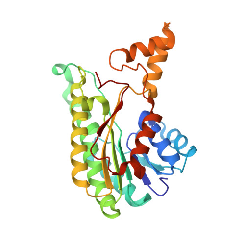
5YSS - PubMed Abstract:
Biocatalytic cyclization is highly desirable for efficient synthesis of biologically derived chemical substances, such as the commodity chemicals ε-caprolactam and δ-valerolactam. To identify biocatalysts in lactam biosynthesis, we develop a caprolactam-detecting genetic enzyme screening system (CL-GESS). The Alcaligenes faecalis regulatory protein NitR is adopted for the highly specific detection of lactam compounds against lactam biosynthetic intermediates. We further systematically optimize the genetic components of the CL-GESS to enhance sensitivity, achieving 10-fold improvement. Using this highly sensitive GESS, we screen marine metagenomes and find an enzyme that cyclizes ω-amino fatty acids to lactam. Moreover, we determine the X-ray crystal structure and catalytic residues based on mutational analysis of the cyclase. The cyclase is also used as a helper enzyme to sense intracellular ω-amino fatty acids. We expect this simple and accurate biosensor to have wide-ranging applications in rapid screening of new lactam-synthesizing enzymes and metabolic engineering for lactam bio-production.
- Synthetic Biology and Bioengineering Research Center, KRIBB, Daejeon, 34141, Republic of Korea.
Organizational Affiliation: