5YS3

1.8 angstrom crystal structure of Succinate-Acetate Permease from Citrobacter koseri

  • Classification: TRANSPORT PROTEIN
  • Organism(s): Citrobacter koseri ATCC BAA-895
  • Expression System: Escherichia coli
  • Mutation(s): No 
  • Membrane Protein: Yes  OPMPDBTMMemProtMD

  • Deposited: 2017-11-13 Released: 2018-04-25 
  • Deposition Author(s): Qiu, B., Liao, J.
  • Funding Organization(s): Ministry of Science and Technology of China, National Science and Technology Major Project, National Key Scientific Instrument and Equipment Development Program, Joint Fund of the National Natural Science Foundation of China and the Israel Science Foundation Research Program, China Post-Doctoral Science Foundation

Experimental Data Snapshot

  • Method: X-RAY DIFFRACTION
  • Resolution: 1.82 Å
  • R-Value Free: 0.199 
  • R-Value Work: 0.160 
  • R-Value Observed: 0.162 

wwPDB Validation   3D Report Full Report


Ligand Structure Quality Assessment 


This is version 1.2 of the entry. See complete history


Literature

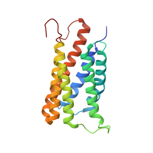
Succinate-acetate permease from Citrobacter koseri is an anion channel that unidirectionally translocates acetate

Qiu, B.Xia, B.Zhou, Q.Lu, Y.He, M.Hasegawa, K.Ma, Z.Zhang, F.Gu, L.Mao, Q.Wang, F.Zhao, S.Gao, Z.Liao, J.

(2018) Cell Res 28: 644-654

  • DOI: https://doi.org/10.1038/s41422-018-0032-8
  • Primary Citation of Related Structures:  
    5YS3, 5YS8

  • PubMed Abstract: 

    Acetate is an important metabolite in metabolism and cell signaling. Succinate-Acetate Permease (SatP) superfamily proteins are known to be responsible for acetate transport across membranes, but the nature of this transport remains unknown. Here, we show that the SatP homolog from Citrobacter koseri (SatP_Ck) is an anion channel that can unidirectionally translocate acetate at rates of the order of ~10 7  ions/s. Crystal structures of SatP_Ck in complex with multiple acetates at 1.8 Å reveal that the acetate pathway consists of four acetate-binding sites aligned in a single file that are interrupted by three hydrophobic constrictions. The bound acetates at the four sites are each orientated differently. The acetate at the cytoplasmic vestibule is partially dehydrated, whereas those in the main pore body are fully dehydrated. Aromatic residues within the substrate pathway may coordinate translocation of acetates via anion-π interactions. SatP_Ck reveals a new type of selective anion channel and provides a structural and functional template for understanding organic anion transport.


  • Organizational Affiliation

    School of Life Science and Technology, ShanghaiTech University, Shanghai, 201210, China.


Macromolecules
Find similar proteins by:  (by identity cutoff)  |  3D Structure
Entity ID: 1
MoleculeChains Sequence LengthOrganismDetailsImage
Succinate-Acetate Permease
A, B, C
192Citrobacter koseri ATCC BAA-895Mutation(s): 0 
Gene Names: CKO_03375
Membrane Entity: Yes 
UniProt
Find proteins for A8ALU5 (Citrobacter koseri (strain ATCC BAA-895 / CDC 4225-83 / SGSC4696))
Explore A8ALU5 
Go to UniProtKB:  A8ALU5
Entity Groups  
Sequence Clusters30% Identity50% Identity70% Identity90% Identity95% Identity100% Identity
UniProt GroupA8ALU5
Sequence Annotations
Expand
  • Reference Sequence
Small Molecules
Ligands 3 Unique
IDChains Name / Formula / InChI Key2D Diagram3D Interactions
78M
Query on 78M

Download Ideal Coordinates CCD File 
AA [auth B]
BA [auth B]
CA [auth B]
LA [auth C]
N [auth A]
AA [auth B],
BA [auth B],
CA [auth B],
LA [auth C],
N [auth A],
O [auth A],
Z [auth B]
(2S)-2,3-DIHYDROXYPROPYL(7Z)-PENTADEC-7-ENOATE
C18 H34 O4
BJMLBVHMHXYQFS-JJEJIETFSA-N
PLM
Query on PLM

Download Ideal Coordinates CCD File 
J [auth A]
JA [auth C]
K [auth A]
KA [auth C]
L [auth A]
J [auth A],
JA [auth C],
K [auth A],
KA [auth C],
L [auth A],
M [auth A],
V [auth B],
W [auth B],
X [auth B],
Y [auth B]
PALMITIC ACID
C16 H32 O2
IPCSVZSSVZVIGE-UHFFFAOYSA-N
ACT
Query on ACT

Download Ideal Coordinates CCD File 
D [auth A]
DA [auth C]
E [auth A]
EA [auth C]
F [auth A]
D [auth A],
DA [auth C],
E [auth A],
EA [auth C],
F [auth A],
FA [auth C],
G [auth A],
GA [auth C],
H [auth A],
HA [auth C],
I [auth A],
IA [auth C],
P [auth B],
Q [auth B],
R [auth B],
S [auth B],
T [auth B],
U [auth B]
ACETATE ION
C2 H3 O2
QTBSBXVTEAMEQO-UHFFFAOYSA-M
Experimental Data & Validation

Experimental Data

  • Method: X-RAY DIFFRACTION
  • Resolution: 1.82 Å
  • R-Value Free: 0.199 
  • R-Value Work: 0.160 
  • R-Value Observed: 0.162 
  • Space Group: P 42
Unit Cell:
Length ( Å )Angle ( ˚ )
a = 79.456α = 90
b = 79.456β = 90
c = 89.099γ = 90
Software Package:
Software NamePurpose
PHENIXrefinement
HKL-2000data reduction
XDSdata reduction
HKL-2000data scaling
SHELXDphasing

Structure Validation

View Full Validation Report


Ligand Structure Quality Assessment 


Entry History & Funding Information

Deposition Data


Funding OrganizationLocationGrant Number
Ministry of Science and Technology of ChinaChina2017YFA0504800
National Science and Technology Major ProjectChina2017ZX09101005-003-003
National Key Scientific Instrument and Equipment Development ProgramChina2012YQ03026010
Joint Fund of the National Natural Science Foundation of China and the Israel Science Foundation Research ProgramChina8146114802
China Post-Doctoral Science FoundationChina2016M600344

Revision History  (Full details and data files)

  • Version 1.0: 2018-04-25
    Type: Initial release
  • Version 1.1: 2018-06-20
    Changes: Data collection, Database references
  • Version 1.2: 2024-03-27
    Changes: Data collection, Database references