5XVD | pdb_00005xvd

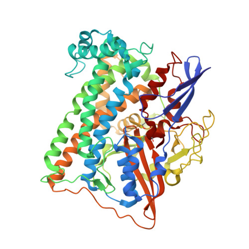
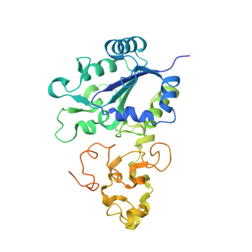
[NiFe]-hydrogenase (Hyb-type) from Citrobacter sp. S-77 in an air-oxidized condition


Experimental Data Snapshot

  • Method: X-RAY DIFFRACTION
  • Resolution: 1.57 Å
  • R-Value Free: 
    0.166 (Depositor), 0.166 (DCC) 
  • R-Value Work: 
    0.121 (Depositor), 0.121 (DCC) 
  • R-Value Observed: 
    0.123 (Depositor) 

wwPDB Validation 3D Report Full Report

Validation slider image for 5XVD

This is version 1.3 of the entry. See complete history. 

Literature

Redox-dependent conformational changes of a proximal [4Fe-4S] cluster in Hyb-type [NiFe]-hydrogenase to protect the active site from O2.

Noor, N.D.M., Matsuura, H., Nishikawa, K., Tai, H., Hirota, S., Kim, J., Kang, J., Tateno, M., Yoon, K.S., Ogo, S., Kubota, S., Shomura, Y., Higuchi, Y.

(2018) Chem Commun (Camb) 54: 12385-12388

  • DOI: https://doi.org/10.1039/c8cc06261g
  • Primary Citation Related Structures: 
    5XVB, 5XVC, 5XVD

  • PubMed Abstract: 

    Citrobacter sp. S-77 [NiFe]-hydrogenase harbors a standard [4Fe-4S] cluster proximal to the Ni-Fe active site. The presence of relocatable water molecules and a flexible aspartate enables the [4Fe-4S] to display redox-dependent conformational changes. These structural features are proposed to be the key aspects that protect the active site from O2 attack.


  • Organizational Affiliation: 
    • Department of Life Science, Graduate School of Life Science, University of Hyogo, 3-2-1 Koto, Kamigori-cho, Ako-gun, Hyogo 678-1297, Japan. hig@sci.u-hyogo.ac.jp.

Macromolecule Content 

  • Total Structure Weight: 197.22 kDa 
  • Atom Count: 14,075 
  • Modeled Residue Count: 1,640 
  • Deposited Residue Count: 1,774 
  • Unique protein chains: 2

Macromolecules

Find similar proteins by:|  3D Structure
Entity ID: 1
MoleculeChains  Sequence LengthOrganismDetailsImage
[NiFe]-hydrogenase 2 large subunitA [auth L],
C [auth M]
552Citrobacter sp. S-77Mutation(s): 0 
EC: 1.12.99.6
UniProt
Find proteins for A0A3B6UEQ1 (Citrobacter sp. S-77)
Explore A0A3B6UEQ1 
Go to UniProtKB:  A0A3B6UEQ1
Entity Groups
Sequence Clusters30% Identity50% Identity70% Identity90% Identity95% Identity100% Identity
UniProt GroupA0A3B6UEQ1
Sequence Annotations
Expand
Reference Sequence
Find similar proteins by:|  3D Structure
Entity ID: 2
MoleculeChains  Sequence LengthOrganismDetailsImage
Hydrogenase-2 small chainB [auth S],
D [auth T]
335Citrobacter sp. MGH106Mutation(s): 0 
EC: 1.12.99.6
UniProt
Find proteins for P69741 (Escherichia coli (strain K12))
Explore P69741 
Go to UniProtKB:  P69741
Entity Groups
Sequence Clusters30% Identity50% Identity70% Identity90% Identity95% Identity100% Identity
UniProt GroupP69741
Sequence Annotations
Expand
Reference Sequence

Small Molecules

Ligands 7 Unique
IDChains Name / Formula / InChI Key2D Diagram3D Interactions
8JU

Query on 8JU


Download:Ideal Coordinates CCD File
M [auth S],
U [auth T]
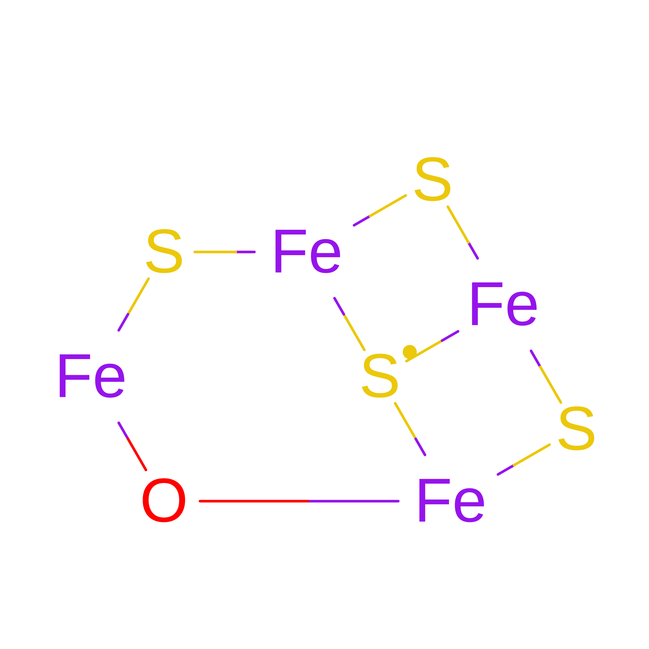
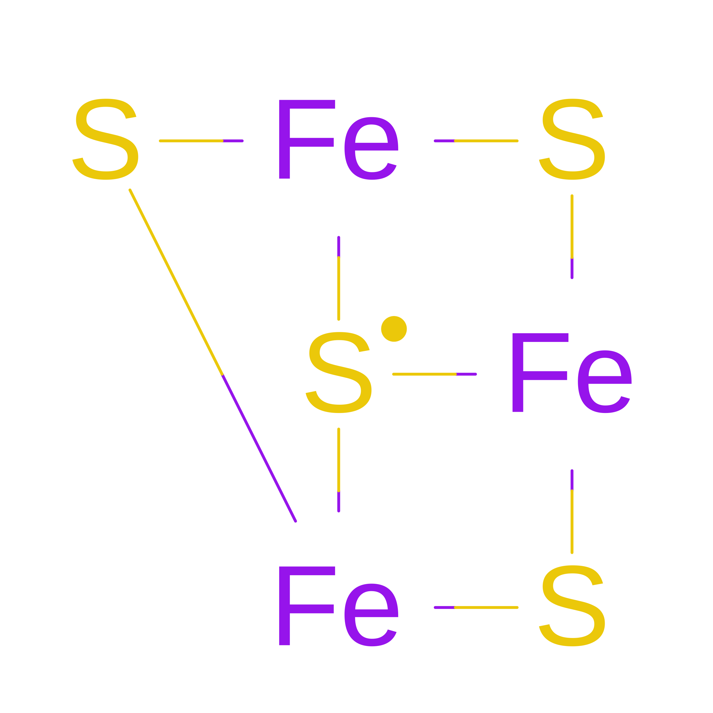
FE4-S4-O CLUSTER
Fe4 O S4
RAQKYMZLKKCSFU-UHFFFAOYSA-N
SF4

Query on SF4


Download:Ideal Coordinates CCD File
J [auth S],
L [auth S],
R [auth T],
T
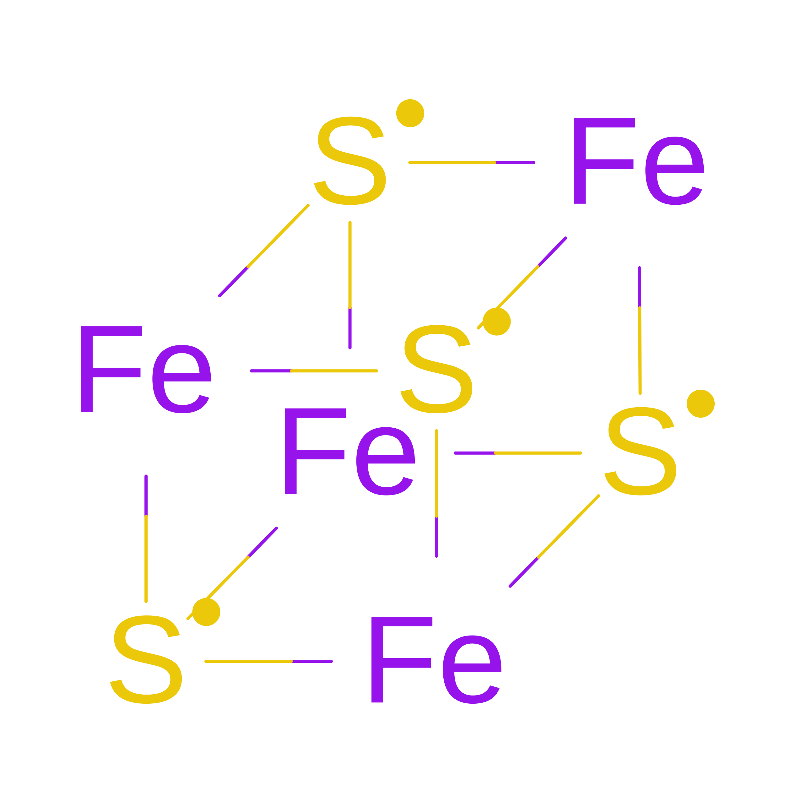
IRON/SULFUR CLUSTER
Fe4 S4
LJBDFODJNLIPKO-UHFFFAOYSA-N
F3S

Query on F3S


Download:Ideal Coordinates CCD File
K [auth S],
S [auth T]
FE3-S4 CLUSTER
Fe3 S4
FCXHZBQOKRZXKS-UHFFFAOYSA-N
NFV

Query on NFV


Download:Ideal Coordinates CCD File
F [auth L],
P [auth M]
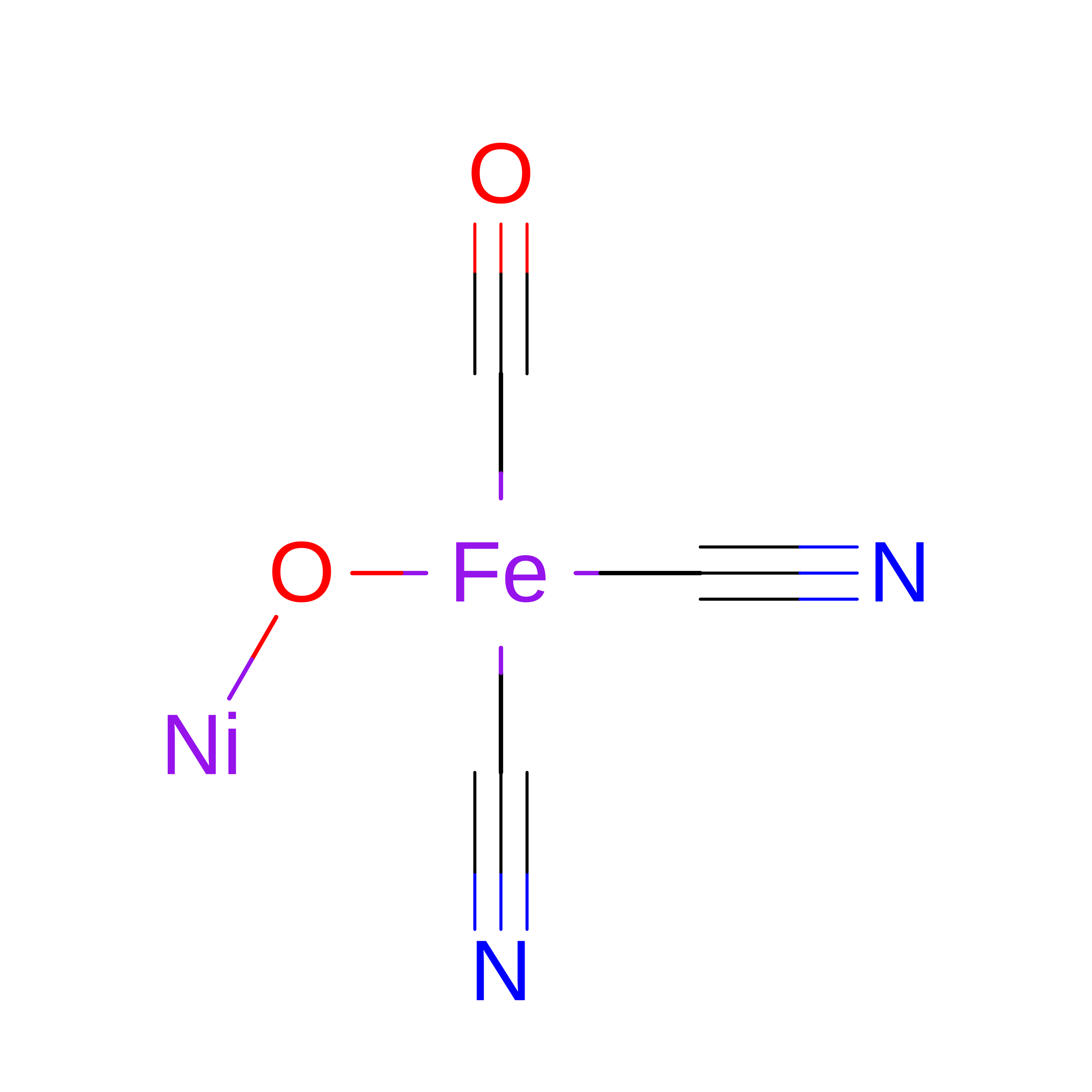
NI-FE OXIDIZED ACTIVE CENTER
C3 Fe N2 Ni O2
MPQMGFDSXFFIQL-UHFFFAOYSA-N
GOL

Query on GOL


Download:Ideal Coordinates CCD File
G [auth L],
H [auth L],
N [auth S],
V [auth T]
GLYCEROL
C3 H8 O3
PEDCQBHIVMGVHV-UHFFFAOYSA-N
MG

Query on MG


Download:Ideal Coordinates CCD File
E [auth L],
O [auth M]
MAGNESIUM ION
Mg
JLVVSXFLKOJNIY-UHFFFAOYSA-N
NA

Query on NA


Download:Ideal Coordinates CCD File
I [auth L],
Q [auth M]
SODIUM ION
Na
FKNQFGJONOIPTF-UHFFFAOYSA-N
Modified Residues  1 Unique
IDChains TypeFormula2D DiagramParent
CSO
Query on CSO
A [auth L],
C [auth M]
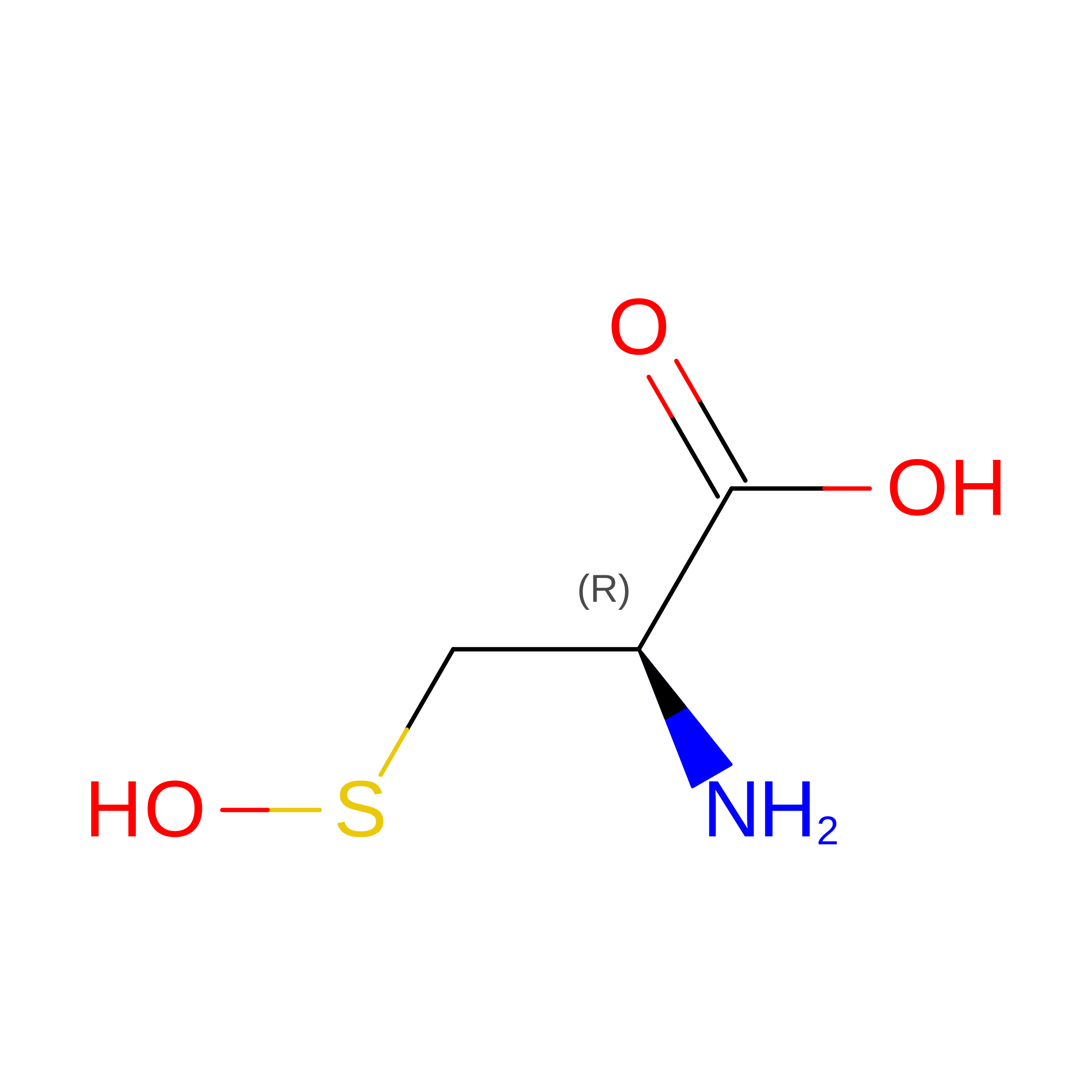
L-PEPTIDE LINKINGC3 H7 N O3 SCYS

Experimental Data & Validation

Experimental Data

  • Method: X-RAY DIFFRACTION
  • Resolution: 1.57 Å
  • R-Value Free:  0.166 (Depositor), 0.166 (DCC) 
  • R-Value Work:  0.121 (Depositor), 0.121 (DCC) 
  • R-Value Observed: 0.123 (Depositor) 
Space Group: P 1 21 1
Unit Cell:
Length ( Å )Angle ( ˚ )
a = 63.94α = 90
b = 118.98β = 100.57
c = 96.81γ = 90
Software Package:
Software NamePurpose
PHENIXrefinement
XDSdata reduction
XSCALEdata scaling
PHASERphasing

Structure Validation

View Full Validation Report


Entry History 

Deposition Data

Revision History  (Full details and data files)

  • Version 1.0: 2018-06-27
    Type: Initial release
  • Version 1.1: 2019-07-10
    Changes: Data collection, Database references, Structure summary
  • Version 1.2: 2024-10-23
    Changes: Data collection, Database references, Derived calculations, Structure summary
  • Version 1.3: 2026-08-12
    Type: Remediation
    Reason: Metalloprotein remediation
    Changes: Derived calculations, Structure summary