5VJ9

Guanidine-II riboswitch P2 hairpin dimer from Pseudomonas aeruginosa

  • Classification: RNA
  • Organism(s): Pseudomonas aeruginosa
  • Mutation(s): No 

  • Deposited: 2017-04-19 Released: 2017-06-28 
  • Deposition Author(s): Reiss, C.W., Strobel, S.A.
  • Funding Organization(s): National Institutes of Health/National Institute of General Medical Sciences (NIH/NIGMS)

Experimental Data Snapshot

  • Method: X-RAY DIFFRACTION
  • Resolution: 1.57 Å
  • R-Value Free: 0.234 
  • R-Value Work: 0.192 
  • R-Value Observed: 0.194 

wwPDB Validation   3D Report Full Report


Ligand Structure Quality Assessment 


This is version 1.4 of the entry. See complete history


Literature

Structural basis for ligand binding to the guanidine-II riboswitch.

Reiss, C.W.Strobel, S.A.

(2017) RNA 23: 1338-1343

  • DOI: https://doi.org/10.1261/rna.061804.117
  • Primary Citation of Related Structures:  
    5VJ9, 5VJB

  • PubMed Abstract: 

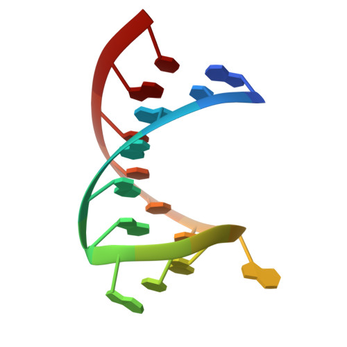
    The guanidine-II riboswitch, also known as mini-ykkC , is a conserved mRNA element with more than 800 examples in bacteria. It consists of two stem-loops capped by identical, conserved tetraloops that are separated by a linker region of variable length and sequence. Like the guanidine-I riboswitch, it controls the expression of guanidine carboxylases and SugE-like genes. The guanidine-II riboswitch specifically binds free guanidinium cations and functions as a translationally controlled on-switch. Here we report the structure of a P2 stem-loop from the Pseudomonas aeruginosa guanidine-II riboswitch aptamer bound to guanidine at 1.57 Å resolution. The hairpins dimerize via the conserved tetraloop, which also contains the binding pocket. Two guanidinium molecules bind near the dimerization interface, one in each tetraloop. The guanidinium cation is engaged in extensive hydrogen bonding to the RNA. Contacts include the Hoogsteen face of a guanine base and three nonbridging phosphate oxygens. Cation-π interactions and ionic interactions also stabilize ligand binding. The guanidine-II riboswitch utilizes the same recognition strategies as the guanidine-I riboswitch while adopting an entirely different and much smaller RNA fold.


  • Organizational Affiliation

    Department of Molecular Biophysics and Biochemistry, Chemical Biology Institute, Yale University, West Haven, Connecticut 06516, USA.


Macromolecules

Find similar nucleic acids by:  Sequence   |   3D Structure  

Entity ID: 1
MoleculeChains LengthOrganismImage
RNA (5'-R(*GP*CP*GP*GP*GP*GP*AP*CP*GP*AP*CP*CP*CP*UP*GP*C)-3')
A, B, C, D
16Pseudomonas aeruginosa
Sequence Annotations
Expand
  • Reference Sequence
Small Molecules
Ligands 4 Unique
IDChains Name / Formula / InChI Key2D Diagram3D Interactions
SPM
Query on SPM

Download Ideal Coordinates CCD File 
F [auth A],
I [auth B],
J [auth B],
L [auth C],
N [auth D]
SPERMINE
C10 H26 N4
PFNFFQXMRSDOHW-UHFFFAOYSA-N
GAI
Query on GAI

Download Ideal Coordinates CCD File 
E [auth A],
G [auth B],
K [auth C],
M [auth D]
GUANIDINE
C H5 N3
ZRALSGWEFCBTJO-UHFFFAOYSA-N
K
Query on K

Download Ideal Coordinates CCD File 
O [auth D]POTASSIUM ION
K
NPYPAHLBTDXSSS-UHFFFAOYSA-N
MG
Query on MG

Download Ideal Coordinates CCD File 
H [auth B]MAGNESIUM ION
Mg
JLVVSXFLKOJNIY-UHFFFAOYSA-N
Experimental Data & Validation

Experimental Data

  • Method: X-RAY DIFFRACTION
  • Resolution: 1.57 Å
  • R-Value Free: 0.234 
  • R-Value Work: 0.192 
  • R-Value Observed: 0.194 
  • Space Group: P 21 21 21
Unit Cell:
Length ( Å )Angle ( ˚ )
a = 50.252α = 90
b = 60.664β = 90
c = 72.308γ = 90
Software Package:
Software NamePurpose
REFMACrefinement
HKL-2000data reduction
SCALEPACKdata scaling
Cootmodel building
PDB_EXTRACTdata extraction

Structure Validation

View Full Validation Report


Ligand Structure Quality Assessment 


Entry History & Funding Information

Deposition Data


Funding OrganizationLocationGrant Number
National Institutes of Health/National Institute of General Medical Sciences (NIH/NIGMS)United StatesGM022778

Revision History  (Full details and data files)

  • Version 1.0: 2017-06-28
    Type: Initial release
  • Version 1.1: 2017-08-30
    Changes: Database references
  • Version 1.2: 2017-09-13
    Changes: Author supporting evidence
  • Version 1.3: 2020-01-01
    Changes: Author supporting evidence
  • Version 1.4: 2024-03-13
    Changes: Data collection, Database references, Derived calculations